Image of the Month February 2025: Congenital Absence of the Pericardium: A Rare Incidental Finding with Key Imaging Insights

Author

Dr Chary Duraikannu

Consultant Radiologist, Ninewells Hospital and Medical School, NHS Tayside, Dundee, UK.


Case history

A 54-year-old male with a long-standing murmur presents for evaluation. A prior transesophageal echocardiogram revealed no septal defects but noted a slightly abnormal appearance of the mitral valve. The patient recently experienced a collapse and has a loop recorder in situ. Previous ECG findings included intraventricular conduction delay, right bundle branch block, and left axis deviation of the QRS complex, with a normal PR interval.

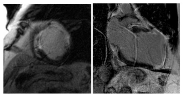
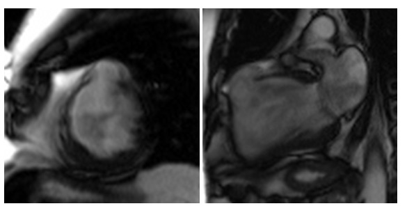
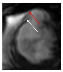
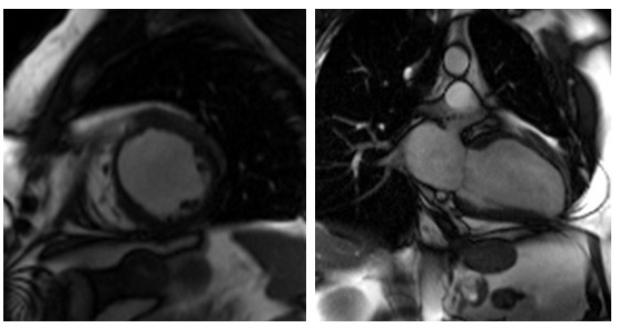
CT and MRI shows complete leftward deviation of heart and midline position of trachea. Interposition of lung tissue noted at aorto-pulmonary window and also discontinuous segment of pericardium in anterior aspect. The heart was otherwise structurally normal, and no intervention was carried out.


Questions

1. Which of the following are imaging features of pericardial agenesis ?

  1. Excessive levorotation
  2. Interposition of lung tissue at aortopulmonary window
  3. Discontinuous segments of the pericardium in anterior aspect
  4. Midline position of the trachea
  5. All of the above

2. Which statement is false ?

  1. Normal pericardium is 1-2mm thick
  2. Pericardium is easily identified along the posterior wall of left ventricle
  3. Pericardial agenesis can be misdiagnosed due to non-specific symptoms
  4. ECG changes include right axis deviation with incomplete RBBB
  5. None of the above

3. Which of the statement is true regarding congenital absence of the pericardium?

  1. Larger defects are typically symptomatic.
  2. Right sided pericardial defects are common than left sided defects.
  3. Risk of herniation of the left atrial appendage in smaller defects.
  4. Majority of patients require surgical intervention.
  5. Cuvier duct regression is not related to pericardial agenesis.


(Answers:e,b,c)


Discussion

Congenital absence of the pericardium is an exceedingly rare anomaly categorized as partial or total defects. Most prevalent are complete left sided defects (70%) followed by complete right sided (17%), complete bilateral (9%) and partial right or left sided (3-4%) [1,2].  Associated congenital anomalies include atrial septal defects, patent ductus arteriosus, mitral valve disease, tetralogy of fallot, and sinus venosus defects with partial anomalous pulmonary venous drainage [3].

In the 5th week of embryonic development pleuropericardial membranes fuse to form pericardium. Failure of this process occurs due to premature regression of ducts of cuvier results in pericardial agenesis [1].

Complete agenesis of pericardium is usually asymptomatic or present with non-specific symptoms. These are mostly incidental during cross sectional imaging performed for other reasons or cardiothoracic surgery [4,5]. However, smaller defects can present with chest pain and dyspnoea when lying on one side and pose risks such as herniation of the left atrial appendage or compression of the left coronary artery [4,6].

Normal pericardium is 1-2 mm in thickness and is usually seen in CT and MRI because of surrounding adipose tissue. Its visualization can be difficult posterior and lateral to left ventricle due to paucity of pericardial fat [7].

Frequently, the electrocardiogram displays bradycardia with right bundle branch block. Additionally, poor R wave progression as well as large P waves may be observed [8].

Diagnosing congenital pericardial defects often relies on indirect signs. Typical findings in cross sectional imaging include excessive levorotation with the left ventricular apex pointing posteriorly, lung interposition at the aorto-pulmonary window and between the base of the heart and the diaphragm, and discontinuous segments of pericardium along the anterior aspect and midline trachea [9,10].

In case of complete bilateral or complete left-sided absence of the pericardium, no treatment is generally indicated [1,11]. Symptomatic patients with partial defects and cardiac chamber herniation should undergo surgery. Treatment options include patch closure of the defect, pericardiectomy, or pericardioplasty [12,13].


Reference:

1.Nasser WK. Congenital diseases of the pericardium. Cardiovasc Clin. 1976;7(3):271-86. PMID: 826317.

2.Klein AL, Abbara S, Agler DA, Appleton CP, Asher CR, Hoit B, Hung J, Garcia MJ, Kronzon I, Oh JK, Rodriguez ER, Schaff HV, Schoenhagen P, Tan CD, White RD. American Society of Echocardiography clinical recommendations for multimodality cardiovascular imaging of patients with pericardial disease: endorsed by the Society for Cardiovascular Magnetic Resonance and Society of Cardiovascular Computed Tomography. J Am Soc Echocardiogr. 2013 Sep;26(9):965-1012.e15. doi: 10.1016/j.echo.2013.06.023. PMID: 23998693.

3.BremerichJ,ReddyGP,HigginsCB.Magneticresonanceimageofcardiacstructure. In: Pohost GM, O’Rourke RA, Berman D, Shah PM, eds. Imaging in Cardiovascular Disease. Philadelphia: Lippincott Williams & Wilkins; 2000. p409, 756.

4.,Bernardinello V, Cipriani A, Perazzolo Marra M, Motta R, Barchitta A. Congenital pericardial agenesis in asymptomatic individuals: tips for the diagnosis. Circ Cardiovasc Imaging 2020;13:e010169.

5.Shiikawa M, Nakahashi K, Endo M, Shiono S. [Congenital Defect of the Pericardium Incidentally Found during Surgery for Lung Cancer:Report of a Case]. Kyobu Geka. 2021 Apr;74(4):308-312. Japanese. PMID: 33831892.

6.Robin E, Ganguly S, Fowler MS. Strangulation of the left atrial appendage
through a congenital partial pericardial defect. Chest 1975;67:354–55

7.Yared K, Baggish AL, Picard MH, Hoffmann U, Hung J. Multimodality imaging of pericardial diseases. JACC Cardiovasc Imaging. 2010 Jun;3(6):650-60. doi: 10.1016/j.jcmg.2010.04.009. PMID: 20541720.

8.Abbas AE, Appleton CP, Liu PT, Sweeney JP. Congenital absence of the pericardium: case presentation and review of literature. Int J Cardiol. 2005 Jan;98(1):21-5. doi: 10.1016/j.ijcard.2003.10.021. PMID: 15676161.

9.Shah AB, Kronzon I. Congenital defects of the pericardium: a review. Eur Heart J Cardiovasc Imaging. 2015 Aug;16(8):821-7. doi: 10.1093/ehjci/jev119. Epub 2015 May 23. PMID: 26003149.

10.Iijima Y, Ishikawa M, Iwai S, Yamagata A, Motono N, Yamagishi S, Koizumi K, Uramoto H. Congenital partial pericardial defect discovered incidentally during surgery for lung cancer: a case report and literature review. BMC Surg. 2021 Dec 31;21(1):447. doi: 10.1186/s12893-021-01453-3. PMID: 34972509; PMCID: PMC8720205.

11.TanakaH,OishiY,MizuguchiY,MiyoshiH,IshimotoT,NagaseNetal.Contribution of the pericardium to left ventricular torsion and regional myocardial function in patients with total absence of the left pericardium. J Am Soc Echocardiogr 2008;21: 268 – 74.

12.Gatzoulis MA, Munk MD, Merchant N, Van Arsdell GS, McCrindle BW, Webb GD. Isolated congenital absence of the pericardium: clinical presentation, diagnosis, and management. Ann Thorac Surg. 2000 Apr;69(4):1209-15. doi: 10.1016/s0003-4975(99)01552-0. PMID: 10800821.

13.Van Son JA, Danielson DG, Schaff HV, Mullany CJ, Julsud PR, Breen JF. Congenital partial and complete absence of the pericardium. Mayo Clin Proc 1993;68:743–7.

Images of the Month January 2025: Incidental finding of congenital absence of the right coronary artery

Authors

Dr Matthew Morgan1, Dr Abbas Ausami1, Dr John Rawlins1

1-University Hospital Southampton NHS Foundation Trust, Southampton, UK


Case history

88yo male presenting with exertional breathlessness and new murmur. Initially investigated with an echo which demonstrated severe aortic stenosis and moderate aortic regurgitation. As part of his work up for TAVI (transcatheter aortic valve implantation), he was referred for coronary angiography and CT coronary angiogram. The patient was found to have a congenital absence of his RCA, with the territory being supplied by a superdominant circumflex artery.

Figure 1 – 3D CTCA reconstruction of the heart from above, demonstrating single coronary origin off the left coronary cusp.Left image: heart and coronary vessels. Right image: coronary vessels with the heart subtracted.

Figure 2 – 3D CTCA reconstruction demonstrating the posterior heart, with a dominant circumflex wrapping round the atrioventricular groove to supply the RCA territory. Left image: heart and coronary vessels. Right image: coronary vessels with the heart subtracted.

Figure 3 – Left image: Coronary angiography demonstrating cannulation of the left mainstem with LAD descending vertically and dominant circumflex wrapping round to supply the RCA territory. Right image: 3D CTCA reconstruction of the coronary arteries demonstrating the same anatomy.


Multiple choice questions

Which of the following congenital abnormalities are associated with absent RCA?

  1. Bicuspid aortic valve
  2. Atrial septal defect
  3. Aortic coarctation
  4. Hypoplastic left heart syndrome
  5. Tetralogy of Fallot


What is the most common symptom associated with absent RCA?

  1. Wheeze
  2. Sudden death
  3. Asymptomatic
  4. Palpitations
  5. Shortness of breath


What is the approximate incidence of absent RCA in the general population?

  1. 0.025%
  2. 0.25%
  3. 25%
  4. 5%
  5. 25%


Answers – 1,3,2


Discussion

Congenital absence of the RCA is a very rare anatomical variant with an estimated incidence of 0.014%-0.066%.1Patients are usually asymptomatic and it is considered a benign pathology, although it can be associated with other cardiac abnormalities such as bicuspid aortic valve, coronary artery fistula and hypertrophic cardiomyopathy.2 None of these were present in this case. Two patterns have been described: L-I pattern where the RCA territory is supplied by an extension of the circumflex artery or LAD (as in this case), and L-II pattern where the RCA territory is supplied by a branch arising from proximal LCA. The L-I pattern appears to be slightly more common and demonstrates a slightly higher incidence of acute MI.3

Previously coronary angiography was considered the gold standard of imaging, however with advances in CTCA, imaging with both modalities is considered useful in giving the most robust assessment of the coronary arteries and their neighbouring structures.3

There is no defined guideline for treatment, which may consist of either conservative or interventional management depending on the severity of coronary atherosclerosis and stenosis. In this case, the symptoms were all felt to be related to the patient’s severe aortic valve disease and he is being worked up for a TAVI.


References:

  1. Zhu XY, Tang XH. Congenital absence of the right coronary artery: A case report. World J Clin Cases2022; 10(34): 12799-12803 [PMID: 36579109 DOI: 12998/wjcc.v10.i34.12799]
  2. Canan, A. and Batra, K., 2022. Superdominant Left Circumflex Artery with Absent Right Coronary Artery. Radiology, 304(2), pp.294-294.
  3. Chen Z, Yan J, Han X, Adhikari BK, Zhang J, Zhang Y, Sun J, Wang Y. Congenital absence of the right coronary artery with acute myocardial infarction: report of two cases and review of the literature. J Int Med Res. 2020 Dec;48(12):300060520971508. doi: 10.1177/0300060520971508. PMID: 33275472; PMCID: PMC7720338.

Image of the month August 2022: Absent left circumflex coronary artery with super-dominant right coronary artery: An extremely rare finding on Computed Tomography Coronary Angiography

Dr Chrysovalantou Nikolaidou1, Dr Andrew D Kelion1

1Radcliffe Department of Medicine, Division of Cardiovascular Medicine, John Radcliffe Hospital, Oxford.


Case history

A 92-year-old woman presented for gated cardiovascular computed tomography for assessment of the aortic annulus and root, coronary arteries and peripheral vascular access prior to Transcatheter Aortic Valve Implantation (TAVI). She had been diagnosed with severe aortic valve stenosis during a routine pre-operative check for treatment of bladder tumours. Her previous medical history included permanent pacemaker implantation for symptomatic second degree atrioventricular block, and paroxysmal atrial fibrillation.

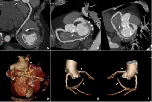
The CT coronary angiogram revealed very unusual coronary anatomy, with complete absence of the left circumflex artery (LCx) (Figure 1, Video 1). There was a huge dominant right coronary artery (RCA), supplying the posterior descending artery and then continuing around the atrio-ventricular groove as a large posterolateral branch to the lateral left ventricular wall. The left coronary artery / left anterior descending artery (LAD) had a normal origin and course (Video 2). There was calcified and mixed plaque disease in both coronary arteries, but no significant luminal stenosis.


Figure 1. Huge right coronary artery supplying the posterior descending artery and then continuing as a large posterolateral branch to the lateral left ventricular wall, as seen on multiplanar reformation (A), maximum intensity projection (B, C) and 3D-volume rendered (D-F) images (white arrows). No left circumflex artery is seen in the left atrio-ventricular groove. The left coronary artery / left anterior descending artery has normal origin and course (white arrowheads), as seen on 3D reconstruction of the coronary artery tree, from the anterior (E) and posterior (F) aspect of the aortic root. Pacemaker wires are also seen in Panel D (white arrowhead).


Video 1: Absence of LCx artery

Video 2: LCA and LAD with a normal anatomy


Questions

  1. Which is the most common congenital coronary artery anomaly?
  1. Congenital absence of the LCx
  2. Origin of the circumflex artery from the RCA or right sinus of Valsalva
  3. Congenital absence of the RCA
  4. Anomalous RCA originating from the LAD or LCx


Answer: B


  1. Which is the most common clinical presentation of coronary artery anomalies?
  1. Chest pain
  2. Sudden cardiac death
  3. Heart failure
  4. Asymptomatic / incidental finding


Answer: D


  1. Congenital absence of the LCx:
  1. Is generally a benign condition
  2. Has been associated with sudden cardiac death
  3. Is a very common congenital coronary artery anomaly
  4. Is an extremely rare anomaly of the coronary arteries


Answers: A, D


Discussion

Congenital coronary artery anomalies may involve the origin, course, and/or structure of the coronary arteries. Their estimated prevalence varies from less than 1% of the general population to 5.8% in the most recent studies using advanced cardiac imaging (1). Most of them are diagnosed incidentally on imaging studies. The most common coronary artery abnormality, excluding separate ostia of the LAD and LCx, is anomalous origin of the circumflex artery from the RCA or right sinus of Valsalva (2). Congenital absence of the LCx is an extremely rare anomaly of the coronary arteries, with an estimated incidence of less than 0.003% to 0.0067% (3, 4). Patients with an absent LCx usually have a large, super-dominant RCA, which supplies blood to the areas of the myocardium usually supplied by the LCx (5). Other concomitant congenital coronary artery anomalies have also been described. Absence of LCx is generally a benign condition. The most common presenting symptom in the majority of cases described in the literature was exertional chest pain. No cases of sudden cardiac death have been reported (3).

Cardiac CT can reliably diagnose congenital coronary artery anomalies, including absence of the LCx, which can be misdiagnosed as ostial occlusion on conventional coronary angiography.


References

  1. Pérez-Pomares JM, de la Pompa JL, Franco D, Henderson D, Ho SY, Houyel L, et al. Congenital coronary artery anomalies: a bridge from embryology to anatomy and pathophysiology—a position statement of the development, anatomy, and pathology ESC Working Group. Cardiovascular Research. 2016;109(2):204-16.
  2. Yuksel S, Meric M, Soylu K, Gulel O, Zengin H, Demircan S, et al. The primary anomalies of coronary artery origin and course: A coronary angiographic analysis of 16,573 patients. Exp Clin Cardiol. 2013;18(2):121-3.
  3. Fugar S, Issac L, Okoh AK, Chedrawy C, Hangouche NE, Yadav N. Congenital Absence of Left Circumflex Artery: A Case Report and Review of the Literature. Case Rep Cardiol. 2017;2017:6579847.
  4. Shaikh SSA, Deshmukh V, Patil V, Khan Z, Singla R, Bansal NO. Congenital Absence of the Left Circumflex Artery With Super-Dominant Right Coronary Artery: Extremely Rare Coronary Anomaly. Cardiol Res. 2018;9(4):264-7.
  5. Rawala MS, Ahmed AS, Iqbal MA, Iqbal A, Budde PK, Rizvi SB. Congenital anomaly of coronary artery: absence of left circumflex artery. Journal of Community Hospital Internal Medicine Perspectives. 2019;9(2):140-2.


Image of the month April 2022- Bean in a bag: a rare case of large pericardial haematoma

Mr Dilliram Adhikari1, Mrs Vimbai Tungwarara1, Dr Badrinathan Chandrasekaran2, Professor Stefan Neubauer1, Dr Chrysovalantou Nikolaidou1

  1. Oxford Centre for Clinical Magnetic Resonance Research, University of Oxford, Oxford
  2. Wiltshire Cardiac Center, Great Western Hospital, Swindon

Case history

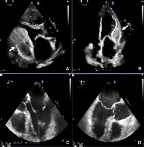
A 82-year-old man, with previous history of bypass surgery (left internal mammary artery to left anterior descending artery, and saphenous venous grafts to obtuse marginal and posterior descending artey) in 2014, was referred for a cardiac magnetic resonance (CMR) scan to further characterise a large pericardial mass seen on echocardiogram (Image 1), which was performed due to newly diagnosed heart failure. Cardiac catheterisation showed patent bypass grafts, and no evidence of pericardial constriction.

Image 1. Transthoracic echocardiogram showing a large pericardial mass on the parasternal long-axis (A) and apical 4-chamber views (B) (arrows). No evidence of the mass on a transoesophageal echocardiogram from May 2017 (C, D).

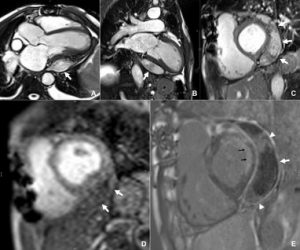
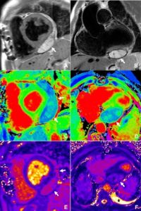
The CMR was performed on a 1.5T Magnetom Avanto-Fit (Siemens, Erlangen, Germany) scanner, using a dedicated protocol for cardiac masses, including cine imaging, tissue characterisation with T1- and T2-weighted imaging and parametric mapping, fat suppression, early and late gadolinium imaging, and rest perfusion imaging. CMR revealed a large, elongated mass (95mm in length x 29 mm in width) within the pericardial space, adjacent to the basal-mid lateral and inferior and the apical inferior left ventricular wall. The mass was well circumscribed with smooth margins and had slightly heterogeneous signal intensity. It did not appear to cross tissue planes and there was no evidence of invasion to the myocardium or extracardiac structures, or compression of the left ventricle. There was no significant pericardial effusion. The mass appeared largely avascular on rest perfusion, early gadolinium enhancement, and late gadolinium enhancement. On late gadolinium imaging, most of the mass showed no enhancement, however, there were small areas of enhancement within the mass and enhancement of its margins (possible fibrous tissue) (Image 2). It had higher signal intensity compared to the myocardium on T1-weighted imaging and T2-weighted imaging and did not demonstrate fat-suppression. Native myocardial T1 values were overall low on T1-mapping (less than 700 ms; normal ShMOLLI myocardial T1 range, 941±23 ms), with pockets of significantly elevated T1 values, while T2 values were significantly elevated on T2-mapping (T2 up to 83 ms; normal myocardial T2 range 48±2 ms at 1.5T in our centre), suggestive of fluid within the mass (Image 3). There was moderate biventricular dilatation and mild biventricular systolic dysfunction, with a moderate-sized myocardial infarction (50-75% wall thickness) in the lateral left ventricular wall.

Image 2. Left ventricular outflow tract (A), vertical long-axis (B) and mid-ventricular short-axis (C) still frames from cine steady-state free precession (SSFP) imaging, showing the large well-circumscribed mass around the lateral and inferior left ventricular wall (arrows). The mass demonstrates minimal contrast uptake on first-pass perfusion imaging (D), only small pockets of enhancement (arrowheads) within the mass and enhancement of its margins (arrow) on free-breathing motion-corrected late gadolinium imaging (E). The black arrows show a previous lateral myocardial infarction.

Image 3. Tissue characterisation of the pericardial mass. Mid-ventricular short-axis T1-weighted image (A) and coronal left ventricular outflow tract T2-weighted image (B) showing the well-demarcated mass with smooth margins, and higher signal intensity compared to the myocardium. T1-mapping short-axis (C) and horizontal long-axis (D) views demonstrating low T1 values of the mass compared to the myocardium. Respective views of the heart on T2-mapping demonstrate high T2 values within the mass. The asterisk shows the small pericardial effusion.


Overall, the findings were in keeping with a benign lesion, which, based on the imaging characteristics, most likely represented organised thrombus/haematoma. A differential diagnosis of secondary or primary cardiac tumour was considered less likely in the absence of invasion across tissue planes and vascular perfusion. The patient denied any history of trauma. Given the absence of compression of cardiac chambers or adjacent structures on CMR and the absence of constrictive physiology on cardiac catheterisation, a conservative management with follow-up imaging was decided. The patient is feeling better on heart failure treatment.


Questions

  1. What are the imaging characteristics of a pericardial cyst on CMR?
  1. They are most commonly seen in the right cardiophrenic angle
  2. They have low to intermediate signal intensity on T1-weighted imaging, and high signal intensity on T2-weighted imaging
  3. They typically enhance after gadolinium administration
  4. They are associated with large pericardial effusion


Answers: A, B


  1. Describe common non-neoplastic pericardial lesions


Answers: pericardial cyst, pericardial diverticula, pericardial haematoma


  1. Which are the CMR tissue characteristics of a pericardial haematoma?
  1. Heterogenous signal intensity, with areas of high T1 and T2 during the subacute phase
  2. Heterogenous signal intensity, with areas of low T1 and high T2 during the subacute phase
  3. Increase in T1 and T2 signal intensity in the chronic phase
  4. Decrease in T1 and T2 signal intensity in the chronic phase


Answers: A, D


Discussion

The normal pericardium consists of two layers, the serous and fibrous pericardium, which appear as a smooth, thin (less than 2 mm), low-intensity curvilinear structure on cine CMR imaging. The pericardial cavity is a small  space, which normally contains between 10 and 50 ml of ultrafiltrate of plasma, and appears as a small rim of fluid around the heart (1). Although pericardial diseases, such as pericarditis or pericardial effusion, are fairly common, pericardial masses are rare, with a prevalence of primary pericardial neoplasms from about 0.001 to 0.007% (2). Pericardial masses can be divided into neoplastic, primary and secondary, and non-neoplastic. The most common benign lesions are pericardial cysts and lipomas. Malignant pericardial masses include mesothelioma, sarcoma, lymphoma and metastatic tumours from the breast, lung and bone marrow (3).

Haemopericardium usually presents acutely after trauma with haemodynamic compromise, however, rare cases of late presentation and progression to constrictive pericarditis and subsequent heart failure have been described. Compared to simple pericardial cysts, haemopericardium is characterised by a heterogeneous high-signal intensity on T1 and T2-weighted CMR imaging in the subacute phase. In the chronic phase, haematomas have a thick rim from hemosiderin deposition and internal foci of varying intensity from calcification, fibrosis, or hemosiderin deposition, the latter resulting in lower signal intensity on native T1 and T2-mapping (4).

CMR can provide information on the size and location of pericardial masses, their extension and relationship to adjacent structures, allowing for assessment of feasibility for surgical resection. Although pathology remains the gold standard for the accurate diagnosis of the type of a mass, CMR can provide crucial non-invasive information on tissue characterisation, and aid the differential diagnosis. CMR can also provide information about pericardial inflammation, adhesions to the myocardium, and evidence of constrictive physiology. In our case, it provided the diagnosis of a benign lesion, most likely pericardial haematoma/thrombus, with no compression of the cardiac chambers, thus helped avoid a re-sternotomy for biopsy of the mass.


References

  1. Bogaert J, Francone M. Cardiovascular magnetic resonance in pericardial diseases. J Cardiovasc Magn Reson. 2009;11(1):14.
  2. Restrepo CS, Vargas D, Ocazionez D, Martínez-Jiménez S, Cuellar SLB, Gutierrez FR. Primary Pericardial Tumors. RadioGraphics. 2013;33(6):1613-30.
  3. Tower-Rader A, Kwon D. Pericardial Masses, Cysts and Diverticula: A Comprehensive Review Using Multimodality Imaging. Prog Cardiovasc Dis. 2017;59(4):389-97.
  4. Watson WD, Ferreira VM, Sayeed R, Rider OJ. Serial Cardiac Magnetic Resonance of an Evolving Subacute Pericardial Hematoma. Circulation: Cardiovascular Imaging. 2019;12(12):e009753.

Image of the Month February 2022: Cement cardiac embolism: A “hard” cause of atrial fibrillation and tricuspid regurgitation

See here for the full CT images

Authors: Samer Alabed1, Boshra Edhayr2 and Annette Johnstone3

1 Radiology Registrar, Sheffield Teaching Hospitals
2 Radiology Registrar, Leeds Teaching Hospitals NHS Trust
3 Consultant Cardiothoracic Radiologist, Leeds Teaching Hospitals NHS Trust


In March 2021 this 72yo Asian female was admitted acutely with a new diagnosis of atrial fibrillation with fast ventricular response. She was started on oral beta blockers, anticoagulated and discharged home. A fortnight later, she was readmitted with increasing breathlessness, and her rate was well controlled at this time.

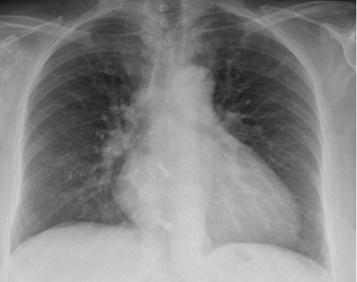
A chest x-ray demonstrated cardiomegaly with upper lobe diversion but no overt pulmonary oedema (Figure 1).

Figure 1: Chest x-ray showing cardiomegaly. A comment was made about calcification in the right hilum and the heart

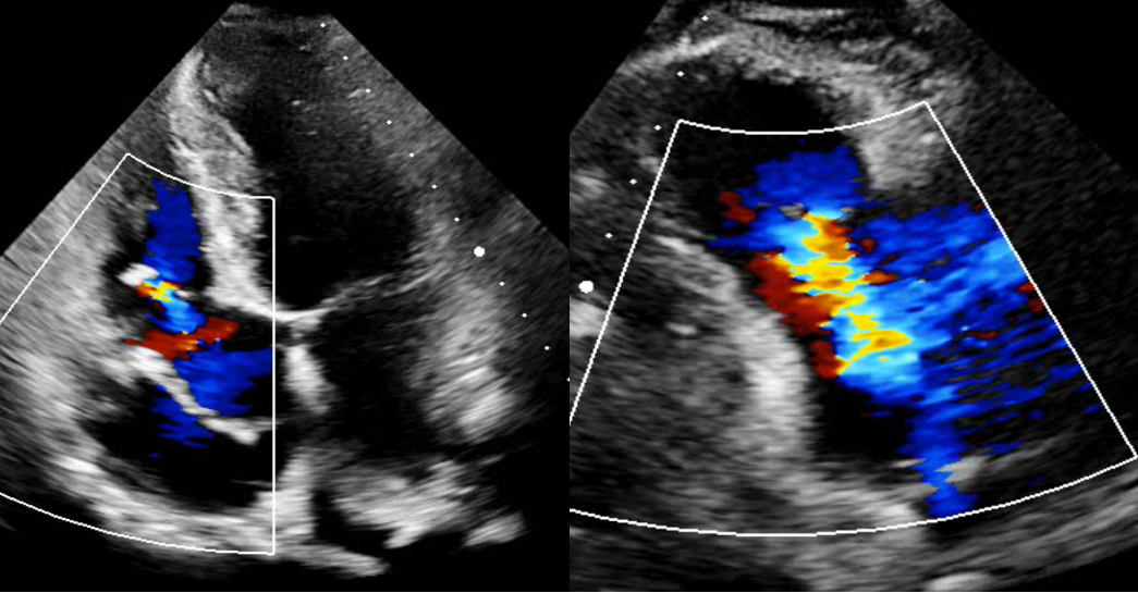
An echocardiogram was subsequently performed which showed normal left ventricular function with septal flattening and dyssynchrony.  The right ventricle was mildly dilated with mildly impaired longitudinal function (TAPSE 14 mm). There was tethering of the anterior tricuspid valve leaflet and complete failure of coaptation of the tricuspid valve, resulting in severe tricuspid regurgitation (Figure 2). There was no significant pericardial effusion.

Figure 2: Echocardiogram demonstrating significant tricuspid regurgitation on the parasternal long axis right ventricular inflow view (right panel). On the apical four chamber view in the left panel, we can see an abnormal structure within the RA attached to the TV leaflet, which was considered to be a prominent Eustachian valve


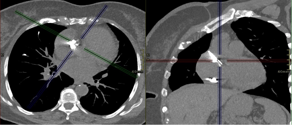
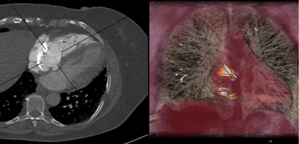
Subsequent HRCT (Figure 3) and CTPA (Figure 4) did not identify an acute pulmonary embolism. However, abnormal high-density material was identified within the right atrium and right atrial appendage with similar material noted within the right upper lobe pulmonary artery and subsegmental divisions of the pulmonary tree. The highly attenuating material within the pulmonary tree demonstrated a tubal and branching configuration. There was no evidence of cardiac perforation and no significant pericardial effusion. Appearances were thought to be compatible with iatrogenic embolization.

Figure 3: HRCT showing high density material in the right atrium. The full CT scan can be accessed through this PACSbin link

Figure 4: CTPA and 3D reconstruction showing high density material in the right atrium and pulmonary arteries (image on the right). The full CT scan can be accessed through this PACSbin link

Reviewing her past medical history, the patient had undergone spinal instrumentation and bilateral knee replacement surgery within the previous year. Interestingly, as her mobility improved, she became more short of breath. A diagnosis of cement embolization was made.

The case was discussed with the regional pulmonary hypertension unit who did not feel pulmonary endarterectomy would be of benefit to this patient. She is currently better, remains anticoagulated and is under regular outpatient review. At the time of writing, she is awaiting surgical discussion regarding the possibility of surgery to remove the material within the right atrium and potential intervention to the tricuspid valve.

Discussion:

Right heart and pulmonary cement embolism is rare and most commonly seen post embolization of polymethyl methacrylate (PMMA) following vertebroplasty procedures or rarely hip replacements. Fresh semi-liquid PMMA can extravasate into the vertebral veins and migrate to the right atrium via the inferior vena-cava. Patients are frequently asymptomatic and cement embolization is usually an incidental finding detected on chest x-ray or cross-sectional imaging. However, very rare cases of fatal cardiac perforation following cement embolization to the heart have been reported.

Treatment options vary, depending on the presentation and symptoms of the individual patient and range from observation and supportive treatment to anticoagulation or embolectomy.

Questions

1. What other types of pulmonary embolism do you know?

Answer: Acute / Chronic thromboembolism, Septic, Fat (trauma), Tumour, Amniotic fluid

2. What is the risk of cement pulmonary embolism after vertebroplasty?

Answer: 5%-7%


3. Which anatomical variants can be found in the atria and can mimic pathology on imaging studies?

Answer: Prominent Eustachian valve, Chiari network, and prominent Crista terminalis in the right atrium; prominent Coumadin ridge in the left atrium; lipomatous hypertrophy of the interatrial septum.


Acknowledgment:
We thank Dr Dominik Schlosshan (Consultant Cardiologist) for providing the echocardiogram images.


References

  1. D’Errico, Stefano, Sara Niballi, and Diana Bonuccelli. 2019. “Fatal Cardiac Perforation and Pulmonary Embolism of Leaked Cement after Percutaneous Vertebroplasty.” Journal of Forensic and Legal Medicine 63 (April): 48–51.
  2. Skjølsvik, Eystein Theodor Ek, Tor Steensrud, Øystein Dahl-Eriksen, Lars Uhlin-Hansen, and Per Ivar Lunde. 2015. “Cardiac Perforation Caused by Cement Embolus after Total Hip Replacement.” Circulation 132 (11): 1047–48.
  3. Zhang, Yi, Xinmei Liu, and Hongsheng Liu. 2022. “Cardiac Perforation Caused by Cement Embolism after Percutaneous Vertebroplasty: A Report of Two Cases.” Orthopaedic Surgery, January. https://doi.org/10.1111/os.13192.

Image of the Month January 2022: Just a benign mass? Value of cardiac magnetic resonance imaging in the diagnosis of cardiac tumours

Authors: Dr Chrysovalantou Nikolaidou1, Dr Marian Lung2, Professor Stefan Neubauer1

  1. Oxford Centre for Clinical Magnetic Resonance Research, University of Oxford, Oxford
  2.  Radcliffe Department of Medicine, John Radcliffe Hospital, Oxford


Case history

A 53-year-old woman, with no significant past medical history, was referred for cardiac MR Imaging (CMR) to further characterise a right atrial mass with imaging characteristics in keeping with cardiac myxoma on echocardiogram (Image 1), which was performed for worsening shortness of breath. Computed tomography revealed extensive lymphadenopathy and multiple lesions within the chest, abdomen, and subcutaneous soft tissue. The blood tests showed elevated white blood cell count with neutrophilia, iron deficiency anaemia, elevated platelet count, and significantly elevated CRP and D-dimers. The provisional diagnosis was cardiac myxoma in the context of possible Carney complex.

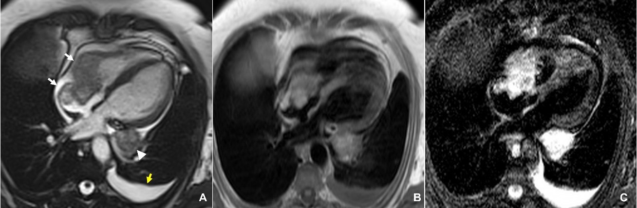
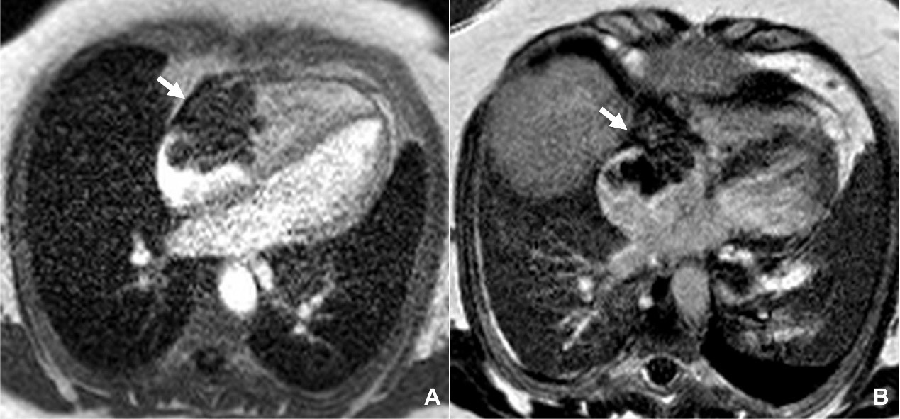
The CMR was performed on a 1.5T Magnetom Avanto (Siemens, Erlangen, Germany) scanner, using a dedicated protocol for cardiac masses, including cine imaging, tissue characterisation with T1- and T2-weighted imaging and parametric mapping, fat suppression, early and late gadolinium imaging, and rest perfusion imaging. CMR revealed a large, irregular-shaped mass (60 x 32 x 45 mm) attached to the lateral right atrial wall and adjacent to the basal-mid inferior and free right ventricular wall, and the right atrioventricular grove. The mass was protruding into and occupying most of the right atrial chamber. Due to the thin right ventricular and right atrial walls, it was difficult to clearly determine the extent of invasion of the mass into these walls. The signal characteristics of the mass were fairly homogeneous, with similar density to the myocardium on T1-weighted imaging and high signal on T2-weighted imaging (Image 2). There was minimal contrast uptake on first-pass perfusion imaging, with minimal heterogeneous enhancement on late gadolinium imaging (Image 3). There was also a second, slightly irregular mass (32 x 25 mm), with similar imaging characteristics, adjacent to the outside of the lateral left atrial wall and left atrioventricular groove.

Overall, the findings were in keeping with a malignant tumour, which, in combination with the widespread disease identified on computed tomography, were most likely to reflect metastatic cardiac involvement from sarcoma or melanoma. The histopathological and molecular investigations on the biopsy samples taken from lymph nodes from the neck and from axilla tissue showed evidence of metastatic melanoma, with little chances of response to systemic treatment. It was clear that the tumour was inoperable, and that the best management was palliative care. The patient died three weeks after the CMR scan.


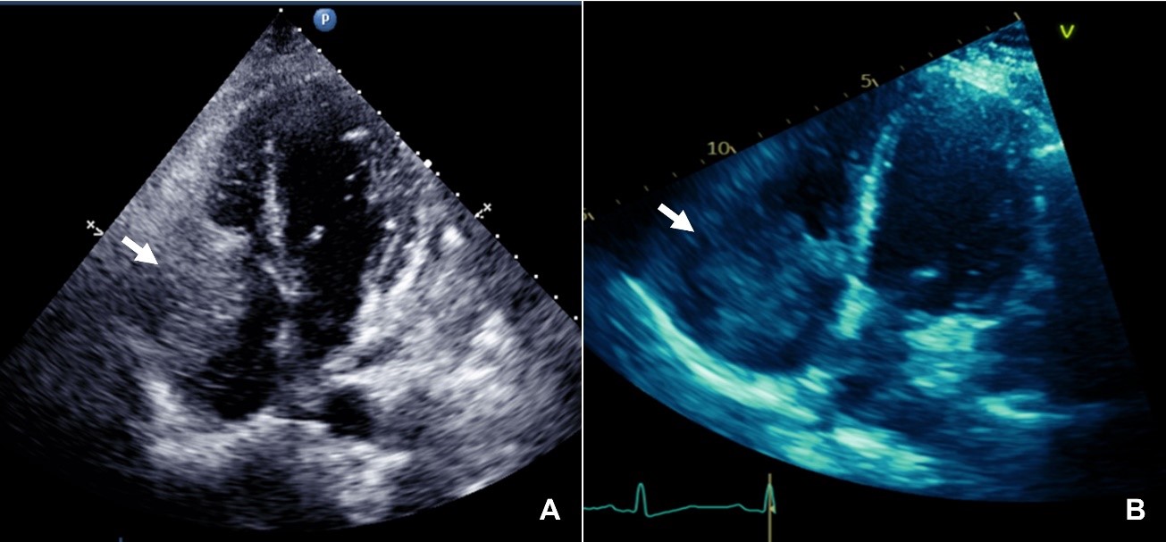
Image 1. Apical four-chamber view (A) and right-ventricular focused view (B), showing a right atrial mass attached to the lateral right atrial wall and protruding into the right ventricle through the tricuspid valve.


Image 2. Horizontal long-axis view of the heart on a still frame from cine steady-state free precession (SSFP) imaging, showing the large irregular right atrial and right ventricular mass (white arrows), a second mass in the basal lateral left ventricular wall (arrowhead), and a small left pleural effusion (yellow arrow) (A). The masses have fairly homogeneous signal, similar to the myocardium on T1-weighted imaging (B), and high signal on T2-weighted imaging. The pleural effusion also appears bright on T2-weighted imaging.


Image 3. Horizontal long-axis view of the heart showing minimal contrast uptake of the mass (white arrow) on first-pass perfusion imaging (A), and minimal heterogeneous enhancement on late gadolinium imaging (B).


Questions

  1. Which of the following statements is true?
  1. The majority of primary cardiac tumours are benign
  2. Primary cardiac tumours are more common than cardiac metastases
  3. The most common benign cardiac tumours are haemangiomas and teratomas
  4. The most common malignant cardiac tumours are sarcomas and lymphomas


Answers: A, D


2. The clinical manifestation of cardiac tumours can include:

  1. Obstruction
  2. Pericardial effusion
  3. Embolic events
  4. All of the above


Answer: D


  1. Which features on CMR imaging are suspicious for a malignant mass?
  1. Large size
  2. Left-sided cardiac location
  3. Tissue inhomogeneity
  4. Low signal on first-pass perfusion imaging


Answers: A, C


Discussion

Cardiac tumours represent a rare pathology, with an estimated prevalence of only 0.002%–0.3% at autopsy. Approximately 75% of primary cardiac tumours are benign, with more than half of these being myxomas. The majority of the malignant primary cardiac tumours are sarcomas and lymphomas. However, metastatic involvement of the heart is more common than primary cardiac tumours, with melanoma, lymphoma, and carcinoma of the lung, breast, and oesophagus being the most frequent initial source. Cardiac tumours can potentially involve any of the cardiac structures and have a diverse clinical presentation, which includes systemic manifestations, symptoms due to mass effect, or embolic events.

While echocardiography remains the first line diagnostic imaging test, CMR has emerged as a powerful diagnostic tool in the diagnosis of many cardiac pathologies, including cardiac masses. CMR can differentiate normal structures (‘pseudotumours’) or cardiac thrombi from abnormal lesions, benign from malignant masses, and provide non-invasive tissue characterisation with the late gadolinium enhancement (LGE) technique, but also with the new T1 and T2-mapping sequences. Imaging features suggestive of a malignant cardiac mass include large size, infiltration of adjacent structures, inhomogeneity on tissue characterisation, right-sided cardiac location, significant contrast uptake on first-pass perfusion imaging, and concomitant pericardial or pleural effusion. However, precise histological assessment is not possible, and pathology remains the gold standard for the accurate diagnosis of the type of a mass, especially in the case of malignant masses. Another advantage of CMR is the ability to assess the vascularity of a mass, its extension, and the relationship to nearby structures, thus helping to guide treatment and assess feasibility of surgical resection.

In conclusion, CMR can reliably evaluate the location, size, and extent of cardiac masses, as well as their invasion across tissue planes. With its unique capability of non-invasive tissue characterisation, CMR can help differentiate between primary and secondary tumours, and provide important clues as to the differential diagnosis.


References

  1. Burazor I, Aviel-Ronen S, Imazio M, et al.Metastatic cardiac tumors: from clinical presentation through diagnosis to treatment. BMC Cancer 2018; 18:  https://doi.org/10.1186/s12885-018-4070-x.
  2. Cardiac tumors. EACVI 3D Echocardiography Box. https://www.escardio.org/Education/Practice-Tools/EACVI-toolboxes/3D-Echo/cardiac-tumors.
  3. Hoffmann U, Globits S, Schima W, et al. Usefulness of magnetic resonance imaging of cardiac and paracardiac masses. Am J Cardiol 2003; 92(7): 890–895.
  4. Motwani M, Kidambi A, Herzog BA, Uddin A, Greenwood JP, Plein S. MR imaging of cardiac tumors and masses: a review of methods and clinical applications. Radiology 2013; 268(1): 26-43. doi: 10.1148/radiol.13121239. PMID: 23793590.
  5. Mousavi N, Cheezum MK, Aghayev A, et al. Assessment of Cardiac Masses by Cardiac Magnetic Resonance Imaging: Histological Correlation and Clinical Outcomes. Journal of the American Heart Association 2019; 8(1): e007829.

Image of the Month December 2021: Closing Holes in Hearts

 Authors: Dr Bronagh P Kelly, Dr Christopher J Lockhart, Dr Mark S Spence, Dr C Owens
Department of Adult Congenital Heart Disease, Belfast Heart Centre, Royal Victoria Hospital, Belfast

Case History:

A 76 year old female presented to the emergency department with hypoxic respiratory failure. She had recently commenced first round of chemotherapy for squamous cell lung cancer and 3 months prior to presentation patient had underwent a right lower lobectomy. Other history included ulcerative colitis, raynaud’s and polymyalgia rheumatica.

Due to increasing oxygen requirements and the need for AIRVO the patient was transferred to the intensive care unit (ICU). Computed tomography pulmonary angiogram (CTPA) demonstrated no evidence of pulmonary embolism/consolidation.

Whilst in ICU, it was observed that saturations improved in the supine position; SaO2 when lying supine was 96% on 4 litres and fell rapidly to 84% on sitting up with the same oxygen administration.

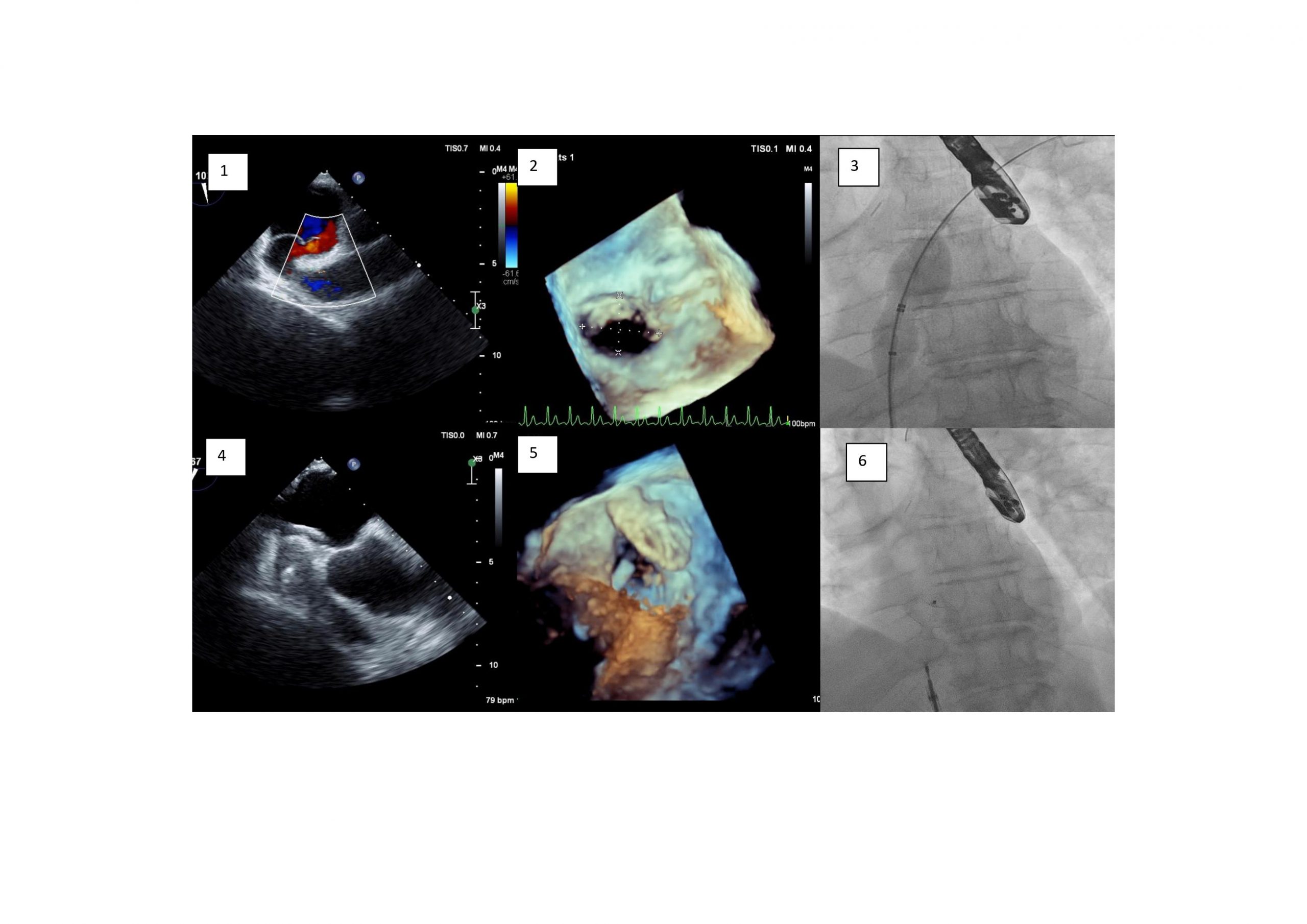
Transthoracic echocardiogram demonstrated a Secundum Atrial Septal Defect.

Bubble echo and transoesophageal echocardiogram demonstrated marked channelling of right atrial flow across a small ASD, demonstrating right to left flow.


The patient proceeded to catheter closure of atrial septal defect using transoesophageal guidance. A sizing balloon was used to occlude the ASD. Saturations rose to 99%. Subsequently a 14 mm Amplatzer septal occluder was deployed using a 9F Torqvue. This resulted in an improvement in the patient’s saturations eliminating the need for additional oxygen supplementation.


Questions

1 . In the presence of an intraatrial communication, what factors precipitate platypnea orthodeoxia syndrome?

  1.  intracardiac shunting
  2. pulmonary shunting
  3. ventilation-perfusion mismatch
  4. all of the above


Answer: D


2.  Which of the following statements are true with regards to indication and method of ASD closure?

  1. An ASD should be closed if there is right ventricular volume overload in the absence of evidence of pulmonary hypertension or left ventricle dysfunction
  2. Surgical closure is the 1st line method for closure of a secundum ASD
  3. Percutaneous device closure is 1st line method of closure of secundum ASD when technically feasible
  4. ASD closure should be considered in patients with suspicion of paradoxical embolism (exclusion of other causes), providing there is absence of PAH and LV disease.
  5. ASD closure is recommended in patients with Eisenmenger physiology.


Answer:

True: A, C, D

False: B, E (Percutaneous device closure is recommended as method of choice for secundim ASD when technically suitable, ASD closure is NOT recommended in patient’s with Eisenmenger physiology)


3.  POS can occur in the presence of intra-atrial shunting either via an ASD or patient foramen ovale (PFO). What are the other indications to close a PFO?

  1. CVA or TIA in patients over the age of 65 in whom paradoxical embolism through patent foramen ovale is considered to be the cause
  2. Migraines
  3. Decompression diving illness
  4. Increased right ventricular volume overload
  5. CVA or TIA in patients under the age of 65 in whom paradoxical embolism through patent foramen ovale is considered to be the cause


Answer: C and E

European Consensus paper 2018 “Perform percutaneous closure of a PFO in carefully selected patients aged from 18 to 65 years with a confirmed cryptogenic stroke, TIA, or systemic embolism and an estimated high probability of a causal role of the PFO as assessed by clinical, anatomical and imaging features” (!)

Decompression diving illness is an indication for PFO closure for high volume divers who wish to continue diving (“). There is insufficient evidence to recommend PFO closure for the indication of migraine.


Discussion:

Platypnoea Orthodeoxia Syndrome (POS) is a rare, undiagnosed, positional dependent phenomenon characterised by hypoxaemia and dyspnoea improves on supine positioning.1

Diagnostic work-up of POS includes transoesophageal echocardiography (TOE) to interrogate for a right to left intracardiac shunt via a patent foramen ovale (PFO) or atrial septal defect (ASD).2

The three main subsets of POS are caused by; intracardiac shunt, pulmonary arteriovenous shunts (eg hepatopulmonary syndrome, hereditary haemorrhagic telangiectasia) or ventilation/perfusions mismatch in the lungs (eg emphysema/COPD, autonomic dysfunction). 3

The pathogenesis of right-to-left atrial shunting with normal intracardiac pressures persists as an area of discussion 4. The median age of patients with POS is in the 7th decade 3

Interatrial right to left shunting is anticipated in the setting of elevated pulmonary pressures, however in cardiac POS, these pressures are usually normal 5.  Therefore, it appears that both an anatomic and a functional component are both required for this shunting to occur, the functional component facilitating the shunting of deoxygenated blood and cause an inverted direction of flow with orthostatism. 3 Although this is not fully understood.

Debilitating dyspnoea secondary to POS can be successfully relieved by closure of the inter-atrial communication6.

Symptomatic improvement is seen in >95% of patients treated with percutaneous closure 5.


References:

  • Knapper, J, Schultz, J. Cardiac Platypnea-Orthodeoxia Syndrome: An Often Unrecognised Malady. ClinCardiol. 2014;37(10): 645-649.
  • Hegland, D, Kunz, G. et al A Hole in the Argument. N Engl J Med. 2005;353: 2385-90.
  • Rodrigues, P, Palma, P, Sousa-pereira, L. Platypnea-Orthodeoxia Syndrome in Review: Defining a new disease?. Cardiology. 2012;123: 15-23
  • Zanchetta, M, Rigatelli, G. A mystery featuring right-to-left shunting despite normal intracardiac pressure. Chest. 2005;128(2): 998-1002
  • Knapper, J, Schultz, J. Cardiac Platypnea-Orthodeoxia Syndrome: An Often Unrecognised Malady. ClinCardiol. 2014;37(10): 645-649.
  • Nassif, M, Lu, H. et al. Neth Heart J. Platypnoea-orthodeoxia syndrome, an underdiagnosed cause of hypoxaemia: four cases and the possible underlying mechanisms. 2015;23: 539-545.
  • Christian Pristipino, Horst Sievert, Fabrizio D’Ascenzo, Jean Louis Mas, Bernhard Meier, Paolo Scacciatella, David Hildick-Smith, Fiorenzo Gaita, Danilo Toni, Paul Kyrle, John Thomson, Genevieve Derumeaux, Eustaquio Onorato, Dirk Sibbing, Peter Germonpré, Sergio Berti, Massimo Chessa, Francesco Bedogni, Dariusz Dudek, Marius Hornung, Jose Zamorano, joint task force of European Association of Percutaneous Cardiovascular Interventions (EAPCI), European Stroke Organisation (ESO), European Heart Rhythm Association (EHRA), European Association for Cardiovascular Imaging (EACVI), Association for European Paediatric and Congenital Cardiology (AEPC), ESC Working group on GUCH, ESC Working group on Thrombosis, European Haematological Society (EHA), European Underwater and Baromedical Society (EUBS), Evidence Synthesis Team, Eapci Scientific Documents and Initiatives Committee, International Experts, European position paper on the management of patients with patent foramen ovale. General approach and left circulation thromboembolism, European Heart Journal, Volume 40, Issue 38, 7 October 2019, Pages 3182–3195, https://doi.org/10.1093/eurheartj/ehy649
  • Landzberg MJ, Khairy P. Indications for the closure of patent foramen ovale. Heart. 2004;90(2):219-224. doi:10.1136/hrt.2003.019315

Image of the Month November 2021 – Computed Tomographic simulation of the anatomical result post transcatheter repair of a sinus venosus defect and partial anomalous pulmonary venous drainage.

Author: Mahon C, Kempny A and Semple T

Royal Brompton and Harefield Hospital, London, United Kingdom.


Educational points:

Three-dimensional volume rendered reformatting with virtual stent implantation can assist in predicting the anatomical result of a catheter-based repair of sinus venosus atrial septal defect (SVASD) and partial anomalous pulmonary venous drainage.


Case Report

A seventy-year-old male with dyspnoea on exertion and new onset pedal oedema was referred for further management of a SVASD. He had presented 10 years prior with atrial fibrillation while undergoing pre-assessment for an elective knee replacement. Echocardiography at the time revealed a SVASD. The patient declined open cardiac surgical repair at the time. He had an uneventful knee replacement and was managed medically from a cardiac perspective. He was referred to our centre for consideration of novel transcatheter repair.

On examination his oxygen saturations were 98% on room air. His body mass index was 29kg/m2, blood pressure 125/80mmHg, resting heart rate 79 beats per minute and irregularly irregular. His jugular venous pressure was elevated with prominent C-V waves. Chest auscultation revealed a grade 2/6 pansystolic murmur and clear lungs. He had a right ventricular heave.

An electrocardiogram (ECG) demonstrated atrial fibrillation at a rate of 72bpm with right bundle branch block and P-pulmonale in leads II, III and AVF.

Repeat transthoracic echocardiography demonstrated a 14 x 20mm SVASD with a left-to-right shunt at rest, severe right ventricle dilation with preserved function and a dilated inferior vena cava with <50% inspiratory collapse.

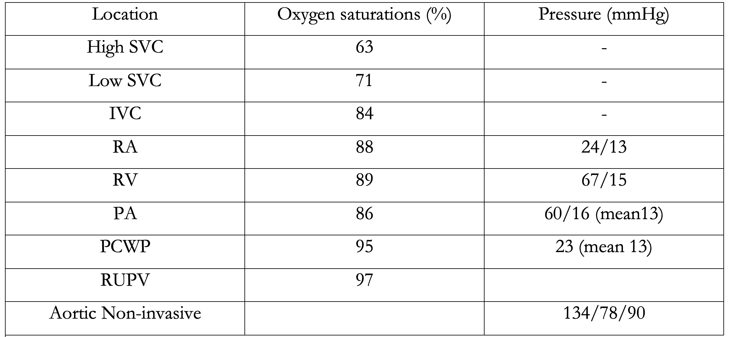
A right heart catheter carried out confirmed left-to-right shunt. Cardiac output was calculated using Fick’s indirect method. The transpulmonary blood flow (Qp) was 9.7L/min, the transsystemic blood flow (Qs) was 4.5 L/min and the Qp/Qs was 2.2. The PVR was normal on invasive assessment at 2.1 woods units (see table 1).

A single-phase ECG triggered high pitch dual source spiral CT acquisition was performed using a weight-based CT angiography protocol. Images were reconstructed at 0.75mm slice thickness at an interval of 0.5mm, and reformatted using Aquarius iNtuition, Terarecon, Durham NC.

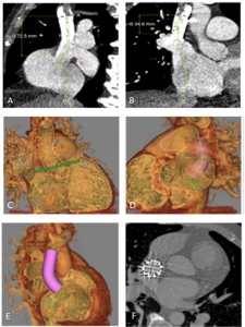
In addition to the SVASD, the CT revealed two PVs anomalously draining to the SVC. The right upper pulmonary vein (RUPV) drained into the SVC immediately above the right atrium (RA) junction. The right middle pulmonary vein (RMPV) drained below the RUPV directly to the RA. The right lower PV and all left PVs drained to the left atrium (LA) (figure 1 a-f). Curved planar reformats suggested that a 7 cm long stent could be used to redirect pulmonary venous return also demonstrated via virtual implantation of a stent (see image 1 images a,e and f).

A balloon test was performed prior to stent insertion. SVC balloon inflation with RUPV and RMPV angiogram was performed to confirm SVASD occlusion, and re-routing of the PVs without obstruction.

The patient underwent successful closure of the SVASD and rerouting of the RUPV and RMPV to the LA (figure 1 g-h).


Table 1: Right heart catheter results

Figure 1 a-b shows a curved planar reformat of the SVC from the level of the azygous to the RA with the SVASD and PAPVD; c shows the three-dimensional volume rendered image of the SVASD; d-e shows the virtual stent and g shows the actual anatomical result post SVASD stent insertion.


Discussion

ASD is a common congenital heart defect with the prevalence of an isolated defect ranging from 0.5 to 2.5 cases per 1,000 live births (1). The resulting shunt depends on the type and size of the defect, the pressures in the RA and LA, the function of the atrioventricular valves and the diastolic properties of the ventricles (2). Many patients are asymptomatic at presentation with an ASD (2).

Closure of an ASD with signs of a significant shunt is associated with improved clinical outcomes as compared to conservative therapy, regardless of age (3). Signs of a significant shunt include right heart dilation or a documented increased Qp/Qs> 1.5. There is increasing evidence to support early ASD closure to improve morbidity (2, 4). The additional volume load from the shunt may lead to right heart dilation and associated progressive tricuspid regurgitation, increased risk of arrhythmia, and right ventricle dysfunction if untreated (2).  In rare cases Eisenmenger’s may occur (2). Our patient was a candidate for intervention with a Qp/Qs of 2.2. He had right heart dilation with preserved function, severe tricuspid regurgitation, and atrial fibrillation in the absence of increased pulmonary vascular resistance on invasive assessment.

Surgical correction is the standard of care and, until recently, was the only treatment option (5,6). The incidences of complications are low with surgical intervention and include the general morbidity of sternotomy and cardiopulmonary bypass as well as specific complications, such as sinus node dysfunction, pulmonary venous obstruction, and occlusion of the SVC (7). Our patient was reluctant for open cardiac surgical intervention, and the novel minimally invasive approach was considered appropriate. The transcatheter approach using a covered stent deployed in the SVC-RA junction was first published by Garg et al in 2014 (8). Since then the technique has been adapted and modified by others (9, 10, 11). The covered stent replaces the deficient posterior wall of the SVC, thereby closing the SVASD redirecting any anomalous PVs into the LA behind the stent (10).

SVASD is well known to be associated with PAPVD (10).   PAPVD can be more complex than a single RUPV or RMPV anomaly entering near the SVC-RA junction or within the RA itself (12). Multiple anomalous PVs or accessory PVs may be present and anomalous PVs on the left side may drain to other locations (7 and 12). Careful selection of patients before transcatheter SVASD closure involves meticulous assessment of the size and position of the PVs relative to the azygous vein and the SVASD prior to stent deployment (7). Both CT and cardiac magnetic resonance imaging can provide a 3-D dataset of the PV anatomy. CT acquisition is quicker with a better special resolution and is currently the preferred modality.

The anomalous PVs need to be directed behind the covered stent to the LA without the stent obstructing the pulmonary venous pathway. Where the junction of the anomalous PV is adjacent to the SVASD, a compromise may be needed between complete closure of the SVASD accompanied by narrowing of the pulmonary venous pathway; leaving a clinically negligible shunt with a widely patent PV; or reconsidering surgery (7). Anomalous PVs that enter high into the SVC may need to remain draining to the SVC. A similar approach is often taken during surgical correction (7). If re-routing is necessary, and/or the risk of PV obstruction is high with stent deployment, then surgery should be considered. In the case presented the CT revealed two anomalous PVs and offered a clear delineation of the PV insertion and anatomic relationship to the SVASD near the RA junction. Superimposition of the stent suggested closure of the SVASD and re-routing of the PAPVD would not cause PV obstruction and could successfully re-route the PVs to the LA. This CT reconstruction with the stent simulation can provide useful information in the decision pathway as to whether to proceed with a transcatheter approach, but cannot replace the requirement for balloon interrogation. The test occlusion will continue to be performed prior to stent deployment, and where there is suggestion of PV obstruction during balloon interrogation of the defect a decision needs to be made whether to continue of abandon the procedure (7).


Conclusion

Careful patient selection before transcatheter SVASD closure and meticulous assessment of the PVs is critical to a successful outcome without complication. CT 3D volume rendered images allow simulation of the stent anchoring positions, aiding decision making for whether to proceed the transcatheter intervention.


References:

  1. van der Linde D, Konings EE, Slager MA, Witsenburg M, Helbing WA, Takkenberg JJ, Roos-Hesselink JW. Birth prevalence of congenital heart disease worldwide: a systematic review and meta-analysis. J Am Coll Cardiol. 2011 Nov 15;58(21):2241-7. doi: 10.1016/j.jacc.2011.08.025. PMID: 22078432.
  2. Kempny A, Gatzoulis MA. Percutaneous repair of sinus venosus ASD: the end of congenital cardiac surgery? EuroIntervention. 2018 Oct 20;14(8):843-845. doi: 10.4244/EIJV14I8A150. PMID: 30339128
  3. Attie F, Rosas M, Granados N, Zabal C, Buendía A, Calderón J. Surgical treatment for secundum atrial septal defects in patients >40 years old. A randomized clinical trial. J Am Coll Cardiol. 2001 Dec;38(7):2035-42. doi: 10.1016/s0735-1097(01)01635-7. PMID: 11738312.
  4. Gatzoulis MA, Freeman MA, Siu SC, Webb GD, Harris L. Atrial arrhythmia after surgical closure of atrial septal defects in adults. N Engl J Med. 1999 Mar 18;340(11):839-46. doi: 10.1056/NEJM199903183401103. PMID: 10080846.
  5. P. Iyer, K. Somanrema, S. Pathak, P.Y. Manjunath, S. Pradhan, S. Krishnan Comparative study of single- and double-patch techniques for sinus venosus atrial septal defect with partial anomalous pulmonary venous connection J Thorac Cardiovasc Surg, 133 (2007), pp. 656-659
  6. D. Stewart, F. Bailliard, A.M. Kelle, C.L. Backer, L. Young, C. Mavroudis Evolving surgical strategy for sinus venosus atrial septal defect: effect on sinus node function and late venous obstructionAnn Thorac Surg, 84 (2007), pp. 1651-1655
  7. Rosenthal E, Qureshi SA, Jones M, Butera G, Sivakumar K, Boudjemline Y, Hijazi ZM, Almaskary S, Ponder RD, Salem MM, Walsh K, Kenny D, Hascoet S, Berman DP, Thomson J, Vettukattil JJ, Zahn EM. Correction of sinus venosus atrial septal defects with the 10 zig covered Cheatham-platinum stent – An international registry. Catheter Cardiovasc Interv. 2021 Jul 1;98(1):128-136. doi: 10.1002/ccd.29750. Epub 2021 May 7. PMID: 33909945.
  8. Garg, H. Tyagi, A.S. Radha Transcatheter closure of sinus venosus atrial septal defect with anomalous drainage of right upper pulmonary vein into superior vena cava—an innovative technique Catheter Cardiovasc Interv, 84 (2014), pp. 473-477
  9. Crystal MA, Vincent JA, Gray WA. The wedding cake solution: A percutaneous correction of a form fruste superior sinus venosus atrial septal defect. Catheter Cardiovasc Interv. 2015 Dec 1;86(7):1204-10. doi: 10.1002/ccd.26031. Epub 2015 May 22. PMID: 26011715. Hansen HJ, Duong P, Jivanji SG, Jones M., Kabir S, Butera . Transcatheter Correction of Superior Sinus Venosus Atrial Septal Defects as an Alternative to Surgical Treatment. JACC 2020 ;75(11)1266-1278
  10. Hansen HJ, Duong P, Jivanji SG, Jones M., Kabir S, Butera . Ttranscatheter Correction of Superior Sinus Venosus Atrial Septal Defects as an Alternative to Surgical Treatment. JACC 2020 ;75(11)1266-1278
  11. Riahi M, Velasco Forte MN, Byrne N, Hermuzi A, Jones M, Baruteau AE, Valverde I, Qureshi SA, Rosenthal E. Early experience of transcatheter correction of superior sinus venosus atrial septal defect with partial anomalous pulmonary venous drainage. EuroIntervention. 2018 Oct 20;14(8):868-876. doi: 10.4244/EIJ-D-18-00304. PMID: 30012542.
  12. Hatipoglu S, Almogheer B, Mahon C, Houshmand G, Uygur B, Giblin GT, Krupickova S, Baksi AJ, Alpendurada F, Prasad SK, Babu-Narayan SV, Gatzoulis MA, Mohiaddin RH, Pennell DJ, Izgi C. Clinical Significance of Partial Anomalous Pulmonary Venous Connections (Isolated and Atrial Septal Defect Associated) Determined by Cardiovascular Magnetic Resonance. Circ Cardiovasc Imaging. 2021 Aug;14(8):e012371. doi: 10.1161/CIRCIMAGING.120.012371. Epub 2021 Aug 13. PMID: 34384233.

Image of the Month October 2021 – Value of an agitated saline bubble contrast ECHO in confirming baffle leaks in post atrial switch for TGA patients

Authors and affiliations

  1. Dr Sindhoora Kotha, Liverpool University Foundation Trust NHS Hospitals
  2. Walter Genco, Manchester Royal Infirmary
  3. Dr Debar Rasoul,  St Helen’s and Knowsley Teaching hospitals NHS trust
  4. Dr Paul Mann, Mid Cheshire hospitals NHS Foundation Trust
  5. Mani Motwani, Manchester Royal Infirmary


Introduction

Complete transposition of great arteries affects about 20-30 of the 100000 livebirths, making it the second commonest cyanotic heart defect (1). The first surgical corrections for complete TGA were developed in 1958 and 1964 and called Mustard and Senning procedures respectively. Both involved redirecting the venous return at the atrial level incorporating baffles (2). Baffle leak and stenosis are well known complications of this kind of repair and can be a major cause of morbidity and mortality in this group of patients (3). This makes early detection of baffle related conundrums very important. Baffle leaks are implicated as a potential risk of stroke acting like a PFO especially in patients with device leads passing through. There are still unanswered questions with regards the best modality of imaging and whether regular screening is recommended to detect baffle leaks at an early stage. We present a case of a young asymptomatic gentleman who underwent a combination of imaging tests to confirm the presence of baffle leak.


Case history

A 33 years old, asymptomatic male, with a background of Complex congenital heart disease in the form of Transposition of great arteries post atrial switch procedure, was referred to the Bubble ECHO clinic, as the Cardiac MR scan was suggestive of a baffle leak via volumetric analysis but was unsuccessful in confirming a shunt visually.

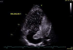
The bubble echo test was performed by injecting agitated saline bubbles via an intra venous cannula in the right arm. The test was found to be negative for a leak at baseline but subsequently suggested significant shunting of bubbles from right to left on increasing intra thoracic pressures by manoeuvres such as coughing and Valsalva (Images 1,2 and 3). By right to left, in this scenario, it meant a leak in the venous baffles causing shunting of blood into the pulmonary baffle.

It is quite difficult to localise the exact position of this leak, but we thought it was most likely from the SVC or the common venous baffle as an injection through the arm would not necessarily opacify the IVC baffle. This patient then went on to have a cardiac MRI scan which suggested a mismatch in Qp:Qs but was unable to demonstrate a shunt on cine imaging.

The bubble echo hence confirmed the presence of the leak and provided a clear visualisation of the baffle leak. Based on the ECHO findings, this gentleman subsequently underwent a formal right heart catheterisation that confirmed a baffle leak for which he then underwent an intervention following an MDT discussion.

Conclusion

Agitated saline contrast studies provide a simple, relatively non-invasive, easily available and reproducible method of routine screening for adults with an atrial switch operation.


Images


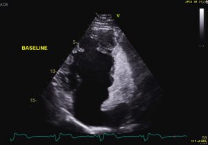
Image 1- Baseline apical 4 chamber view showing the sub pulmonic ventricle opaque with agitated saline bubbles but none entering the systemic side of the circulation


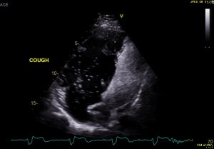
Image 2- Apical 4 chamber view displaying subpulmonic side opaque with agitated saline bubbles and a significant shower of bubbles on coughing seen entering the systemic side (pulmonary baffle and systemic ventricle)

Image 3- Apical 4 chamber view displaying subpulmonic side opaque with agitated saline bubbles and a significant shower of bubbles seen entering the systemic side (pulmonary baffle and systemic ventricle) on performing the Valsalva manoeuvre.

References

1. De Pasquale G et al. High prevalence of baffle leaks in adults after atrial switch operations for transposition of the great arteries. Eur Heart J- cardiovascular imaging. 2017; 18: 531-535
2. Sommer RJ, Hijazi ZM, Rhodes JF. Pathophysiology of Congenital Heart Disease in the Adult: Part III: Complex Congenital Heart Disease. Circulation. 2008: 117: 1340-1350.
3. Wilhelm et al. Accuracy of Imaging Modalities in Detection of Baffle Leaks in Patients Following Atrial Switch Operation. Echocardiography. 2015; 33: 437- 442.

Image of the Month August 2021 – The elusive napkin ring sign

The elusive napkin ring sign

Authors and affiliations

  1. Sai Viswan Thiagarajah, Medical Student, University of Edinburgh
  2. Michelle C Williams, Consultant Radiologist, Centre for Cardiovascular Science, University of Edinburgh, Edinburgh, UK


Case history

A 58-yr old female presented to the emergency department with heavy central chest pain. It was relieved with sublingual glyceryl trinitrate(GTN) and resolved by the time she arrived at the hospital.

She had a previous history of coronary artery disease and suffered a non-ST elevation myocardial infarction (NSTEMI) 1 year previously. An invasive coronary angiogram performed at this time showed severe stenosis in the distal right coronary artery, which was treated with a stent. She had a history of hypertension and a family history of coronary artery disease. She was an ex-smoker with a significant pack year history.

An electrocardiogram (ECG) showed longstanding T wave inversion, but was otherwise unremarkable. High sensitivity troponin on admission was 5 ng/L, and a repeat at 3 hours later was 4 ng/L. She was discharged and followed up with an outpatient computed tomography coronary angiogram (CTCA) which is shown below.

Image

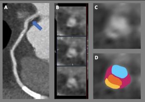
CTCA identified a napkin ring sign in the proximal right coronary artery. Image (A) shows a curved planar reformation of the right coronary artery with an arrow showing the area of the napkin ring sign in the proximal vessel and a patent stent in the distal vessel. (B) shows cross sectional images through the proximal right coronary artery showing three images of the napkin ring sign at 1 mm intervals along the vessel lumen. (C) shows a zoomed in cross sectional image of the napkin ring with (D) showing corresponding labelled components.

Questions & best answers

  1. Which of the following are described as ‘high risk’ features for a plaque seen on CT coronary angiography
    1. Napkin ring sign
    2. Positive vessel remodelling
    3. Spotty calcification
    4. Low attenuation
    5. All of the above

2. Which of the following correctly describes the napkin ring sign?

    1. Calcified plaque with positive vessel remodelling, low attenuation centre and high attenuation peripheral rim
    2. Non-calcified plaque with positive vessel remodelling, low attenuation centre and a high attenuation peripheral rim
    3. Calcified plaque with positive vessel remodelling, high attenuation centre and low attenuation peripherally
    4. Non-calcified plaque with positive vessel remodelling, high attenuation centre and low attenuation peripherally
    5. Calcified plaque with positive remodelling, low attenuation centre, and an isointense peripheral rim.

3. Which of the following is the denotation used in the CAD RADS (Coronary Artery Disease Reporting and Data System) when an adverse plaque feature is seen

    1. CAD RADS 4
    2. CAD RADS 5
    3. Modifier V
    4. Modifier G
    5. Modifier S

Answers

  1. E – all of these have been described as high risk/adverse plaque features
  2. B – In the D shown above, blue represents the lumen, orange represents the low attenuation centre of the napkin ring, pink represents non calcified plaque and yellow represents the high attenuation rim
  3. C – CAD RADS 4 and 5 describe the degree of vessel stenosis, modifier G is for grafts, S is for stents and V is for vulnerable/adverse plaque features


Discussion

CTCA can identify features of high-risk plaques (also called vulnerable plaques, or adverse plaque characteristics) that are thought to be associated with the thin cap fibroatheroma, the histological precursor of ruptured plaques.

Positive vessel remodelling refers to a plaque with the outer vessel diameter > 1.1 times that of the adjacent uninvolved vessel(1). A low attenuation plaque is a non-calcified plaque measuring < 30 Hounsfield units. Spotty calcifications are small, calcified plaques < 3mm in diameter in any direction(2). The napkin ring sign refers to a combination of plaque characteristics where there is a non-calcified plaque with positive remodelling, a low attenuation plaque centre and a higher attenuation peripheral rim, as seen in this case(3). Reporting of these features is recommended in the Coronary Artery Disease Reporting and Data System (CAD RADS), but observer agreement amongst expert readers is only ‘fair’(4).

Several studies have evaluated the usefulness of these vulnerable plaque characteristics for assessing risk of clinical outcomes. Motoyama et al. showed that, those with adverse plaque features were ten times more likely to develop an acute coronary syndrome at 4 year follow up(5). In the PROMISE trial, adverse plaque features was associated with an increased risk of MACE (major adverse cardiovascular events), particularly amongst women and younger patients(6). In the SCOT-HEART trial, adverse plaque characteristics were associated with an increased risk of fatal or non-fatal myocardial infarction, with the greatest risk seen in those with adverse plaques and obstructive coronary artery disease(7). However, in the SCOT-HEART trial adverse plaque characteristics were not an independent predictor of outcomes when controlled for coronary plaque burden assessed with calcium score.

The napkin ring sign has been shown to be a particularly high-risk plaque feature. Maurovich-Horvat et al. showed that the napkin ring sign was associated with histologically advanced atherosclerotic lesions (per plaque sensitivity 36%, specificity 100%)(8). In another study it was also found to be a predictor of subsequent acute coronary syndrome 3 years after follow-up(9). Puchner et al. studied 472 patients with chest pain suspicious of ACS who underwent CTCA(2). The incidence of a napkin ring sign was around ten times greater in those found to have ACS than those without (32.4% vs 3.2%).

However, the napkin ring sign is not a particularly common finding on CTCA. In the SCOT-HEART trial the napkin ring occurred in 0.3% (78 of 26,525) of vessel segments analysed(7). Otsuka et al identified it in 0.4% (45 of 12,727) of segments(9). Uncertainty also remains as to what exactly the napkin ring sign represents – is it focal ulceration, contrast enhancement with the vasovasorum, low density calcification, or something else? The management of these CTCA findings is also uncertain. There are no randomised controlled trials assessing the impact of different management strategies on the progression of these plaques, or subsequent clinical outcomes.


Learning points

  1. Adverse plaque features are common on CTCA but inter-observer ability to recognise their presence is variable
  2. The napkin ring sign is the least common of the adverse plaque features but potentially the most clinically significant
  3. More research is required to understand whether patients with these adverse plaque features on CTCA would benefit from more aggressive management


References

  1. Pundziute G, Schuijf JD, Jukema JW, Decramer I, Sarno G, Vanhoenacker PK, et al. Evaluation of plaque characteristics in acute coronary syndromes: Non-invasive assessment with multi-slice computed tomography and invasive evaluation with intravascular ultrasound radiofrequency data analysis. Eur Heart J [Internet]. 2008 Oct [cited 2021 May 5];29(19):2373–81. Available from: https://pubmed.ncbi.nlm.nih.gov/18682447/
  2. Puchner SB, Liu T, Mayrhofer T, Truong QA, Lee H, Fleg JL, et al. High-risk plaque detected on coronary CT angiography predicts acute coronary syndromes independent of significant stenosis in acute chest pain: Results from the ROMICAT-II trial. J Am Coll Cardiol. 2014 Aug 19;64(7):684–92.
  3. Vulnerable plaque | Radiology Reference Article | Radiopaedia.org [Internet]. [cited 2021 May 17]. Available from: https://radiopaedia.org/articles/vulnerable-plaque?lang=gb
  4. Maroules CD, Hamilton-Craig C, Branch K, Lee J, Cury RC, Maurovich-Horvat P, et al. Coronary artery disease reporting and data system (CAD-RADSTM): Inter-observer agreement for assessment categories and modifiers. J Cardiovasc Comput Tomogr. 2018 Mar 1;12(2):125–30.
  5. Motoyama S, Ito H, Sarai M, Kondo T, Kawai H, Nagahara Y, et al. Plaque characterization by coronary computed tomography angiography and the likelihood of acute coronary events in mid-term follow-up. J Am Coll Cardiol. 2015 Jul 28;66(4):337–46.
  6. Ferencik M, Mayrhofer T, Bittner DO, Emami H, Puchner SB, Lu MT, et al. Use of high-risk coronary atherosclerotic plaque detection for risk stratification of patients with stable chest pain: A secondary analysis of the promise randomized clinical trial. JAMA Cardiol [Internet]. 2018 Feb 1 [cited 2021 Jun 10];3(2):144–52. Available from: https://pubmed.ncbi.nlm.nih.gov/29322167/
  7. Williams MC, Moss AJ, Dweck M, Adamson PD, Alam S, Hunter A, et al. Coronary Artery Plaque Characteristics Associated With Adverse Outcomes in the SCOT-HEART Study. J Am Coll Cardiol [Internet]. 2019 Jan 29 [cited 2021 May 17];73(3):291–301. Available from: /pmc/articles/PMC6342893/
  8. Maurovich-Horvat P, Schlett CL, Alkadhi H, Nakano M, Otsuka F, Stolzmann P, et al. The napkin-ring sign indicates advanced atherosclerotic lesions in coronary CT angiography. JACC Cardiovasc Imaging. 2012 Dec 1;5(12):1243–52.
  9. Otsuka K, Fukuda S, Tanaka A, Nakanishi K, Taguchi H, Yoshikawa J, et al. Napkin-ring sign on coronary CT angiography for the prediction of acute coronary syndrome. JACC Cardiovasc Imaging. 2013 Apr;6(4):448–57.
Our Sponsors