Image of the Month – October 2017 – Significant ischaemic heart disease in a young adult – implementing ESC guidelines in clinical practice

Williams MG1; Harries I2; Kassimis G3; Bucciarelli-Ducci, C1

1Department of Cardiology, Bristol Heart Institute, United Kingdom

2Department of Radiology, Bristol Royal Infirmary, United Kingdom

3Department of Cardiology, Cheltenham General Hospital, United Kingdom

Case report

A previously healthy 34 year old man presented acutely to a district general hospital with chest pain. An electrocardiogram confirmed sinus rhythm with an inferior ST elevation myocardial infarction. His risk factors included a family history of premature coronary artery disease (father, aged 35 years), hypercholesterolaemia (total cholesterol 9 mmol/litre) and elevated body mass index (29 kg/m2). He was taken immediately for a coronary angiogram. This revealed a dominant right coronary artery which was occluded at the posterior descending artery (PDA) (Figure 1a). He also had chronic total occlusions of his circumflex, distal LAD and first diagonal arteries (Figure 1b). Primary percutaneous coronary intervention (PCI) was then performed. Balloon angioplasty of his occluded PDA was done as the culprit lesion and the patient was started on standard secondary prevention medication. Outpatient stress perfusion cardiac magnetic resonance imaging (CMR) was arranged to guide further potential revascularisation strategies.

CMR demonstrated low normal left ventricular systolic function with an ejection fraction of 59%. There was hypokinesia of the mid and apical anterior walls and of the basal inferior wall. Stress images were obtained using a standard adenosine protocol and showed extensive inducible myocardial perfusion defects affecting the basal and mid-anterior, anterolateral, inferolateral walls, mid and apical inferior walls (8 segments in total) (See Figures 2 and 3). There was a small focus of subendocardial late gadolinium enhancement (LGE) of the basal inferior segment (Figure 4).  If technically feasible, revascularisation of the circumflex and left anterior descending territories was advised on prognostic grounds.

The patient underwent elective PCI to his circumflex coronary artery with an excellent result (Figure 5). Technical challenges precluded revascularisation of the diagonal and distal LAD territories and so aggressive secondary prevention and anti-anginal therapy have been commenced.
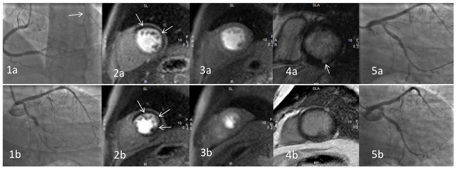
Figure 1. Coronary angiography of the right (1a) and left (1b) coronary arteries; note collateralisation to the chronically occluded circumflex (arrowed)

Figure 2. T1-weighted basal (2a) and mid ventricular (2b) short-axis stress perfusion images demonstrating an extensive inducible perfusion defect (arrowed), predominantly affecting the left anterior descending and left circumflex coronary artery territories.

Figure 3. T1-weighted basal (3a) and mid ventricular (3b) short-axis rest perfusion images demonstrating normal perfusion.

Figure 4. T1-weighted basal (4a) and mid ventricular (4b) short-axis late gadolinium enhancement sequences demonstrating minimal late enhancement in the inferior segment (arrowed).

Figure 5. Percutaneous coronary intervention to the circumflex artery (5a) with an excellent final result (5b).

Multiple choice questions

1. According to current ESC guidelines, what is considered to be a significant ischaemic burden that warrants revascularisation?

A) 5%

B) 10%

C) 15%

D) 20%

E) 25%

Answer B)

Current guidelines suggest that in patients with stable coronary artery disease, a proven large area of ischaemia (≥ 10% of the left ventricle) on non-invasive testing warrants revascularisation on prognostic grounds1. However, recent evidence has suggested that an ischaemic burden of >1.5 segments on cardiac MRI is a strong predictor of poor outcome and this may be reflected in future guidelines2.


2. In patients with multi vessel disease after primary PCI, what level of evidence is quoted by the 2017 ESC STEMI guidelines for the role of cardiac MRI to assess for myocardial ischaemia and viability to guide complete revascularisation?

A) It is not recommended by the ESC

B) I

C) IIa

D) IIb

E) III

Answer D) IIb. The ESC STEMI guidelines (2017)3 state that CMR may be used after primary PCI to assess for ischaemia and viability including in multivessel coronary artery disease.


References

1. Gilles Montalescot (France), Udo Sechtem (Germany), Stephan Achenbach (Germany), Felicita Andreotti (Italy), Chris Arden (UK), Andrzej Budaj (Poland), Raffaele Bugiardini (Italy), Filippo Crea (Italy), Thomas Cuisset (France), Carlo Di Mario (UK), J. Rafael Ferreira (Portugal), Bernard J. Gersh (USA), Anselm K. Gitt (Germany), Jean-Sebastien Hulot (France), Nikolaus Marx (Germany), Lionel H. Opie (South Africa), Matthias Pfisterer (Switzerland), Eva Prescott (Denmark), Frank Ruschitzka (Switzerland), Manel Sabate ́ (Spain), Roxy Senior (UK), David Paul Taggart (UK), Ernst E. van der Wall (Netherlands), Christiaan J.M. Vrints (Belgium). 2013 ESC guidelines on the management of stable coronary artery disease. European Heart Journal (2013) 34, 2949–3003

2. Gabriella Vincenti, Pier Giorgio Masci, Pierre Monney, Tobias Rutz, Sarah Hugelshofer, Mirdita Gaxherri, Olivier Muller, Juan F. Iglesias, Eric Eeckhout, Valentina Lorenzoni, Cyril Pellaton, Christophe Sierro, Juerg Schwitter: Stress Perfusion CMR in Patients With Known and Suspected CAD. JACC: Cardiovascular Imaging May 2017, 10 (5) 526-537; DOI: 10.1016/j.jcmg.2017.02.006

3. Borja Ibanez, Stefan James, Stefan Agewall, Manuel J Antunes, Chiara Bucciarelli-Ducci, Héctor Bueno, Alida L P Caforio, Filippo Crea, John A Goudevenos, Sigrun Halvorsen, Gerhard Hindricks, Adnan Kastrati, Mattie J Lenzen, Eva Prescott, Marco Roffi, Marco Valgimigli, Christoph Varenhorst, Pascal Vranckx, Petr Widimský, ESC Scientific Document Group ; 2017 ESC Guidelines for the management of acute myocardial infarction in patients presenting with ST-segment elevation: The Task Force for the management of acute myocardial infarction in patients presenting with ST-segment elevation of the European Society of Cardiology (ESC), European Heart Journal, ehx393, https://doi.org/10.1093/eurheartj/ehx393
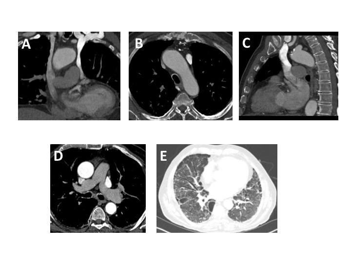
Image of the month – September 2017 – Common systemic venous anomaly with an uncommon configuration

Will Loughborough (ST5 Radiologist), Stephen Lyen (Consultant Cardiothoracic Radiologist)

Affiliations; Bristol Royal Infirmary

Case

An 81 year old male was admitted with severe dyspnoea and hypoxia. A CT pulmonary angiogram (CTPA) was performed to identify a potential pulmonary embolus.

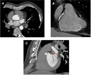
A standard protocol CTPA was performed with intravenous contrast injected via a left arm injection. A bolus tracking acquisition was used with the region of interest (ROI) centered on the main pulmonary artery (MPA). Following contrast administration, there was mild MPA opacification (below the contrast peak required for acquisition trigger). When the aorta started to opacify normally, the acquisition was manually triggered. Upon review of images, it was clear there were both right and left SVCs (figures A and B), with a small brachiocephalic venous connection. The right SVC connected to the right atrium. Unusually, the left SVC drained into the left atrium (figure C) as opposed to the coronary sinus. This explained the unusual contrast haemodynamics during study acquisition, whereby the predominant contrast flow via a left arm injection was through the left SVC into the left atrium. This is a right to left shunt, which had not resulted in any clinically significant haemodynamic consequences and indeed left atrial/ventricular size and left ventricular function were normal on echocardiography. Given this shunt, there was sub-optimal opacification of the pulmonary arteries (figure D), producing a non- diagnostic study for the detection of pulmonary emboli.  A potential option would be attempting the study again with a right sided injection (with a normal drainage into the right atrium), although given the small brachiocephalic venous connection with the left SVC, there would be likely shunting of contrast and potentially, further sub-optimal MPA opacification. A ventilation/perfusion scintigraphy study was not possible due to background Usual Interstitial Pneumonitis (UIP) type pulmonary fibrosis (figure E) and centrilobular and paraseptal emphysema, which would impair results. The clinicians felt this background lung disease plus a co-existing lower respiratory tract infection explained the presenting symptoms so no further imaging was performed.


Figure Legends

Figure A. Coronal CTPA reformat – SVC duplication, with intravenous contrast delivered by left arm injection with opacification seen predominantly in the left SVC.

Figure B. Axial CTPA demonstrating greater opacification of the left-sided SVC, when compared to the right SVC

Figure C. Sagittal CTPA image through the level of the left SVC which is shown to communicate with the left atrium.

Figure D. Axial slice through the level of the MPA – non-diagnostic pulmonary arterial opacification.

Figure E – Axial slice through the lung bases on lung window settings – bronchiectasis and sub-pleural honeycombing with architectural distortion, classical for UIP-type pulmonary fibrosis.

Discussion

The most common variation to the systemic thoracic venous system is a left-sided SVC, occurring in 0.3-0.5% of the population1,2. This occurs alongside a right SVC (‘SVC duplication’) in 90% of cases, but in the remainder, a left-sided SVC occurs in isolation. In 90% cases, a left SVC connects to the coronary sinus (normal systemic venous return from the left subclavian/internal jugular veins into the right atrium)3. However, in 10% cases, as in this case, the left SVC connects directly to the left atrium 4, resulting in a right to left shunt.  The presence of a left-sided SVC has a 5% association with congenital cardiac defects, most commonly an atrio-septal defect 2. A persistent left SVC results from failure of the left anterior cardinal vein to obliterate during early embryological development 2.

Whilst a left-sided SVC connected to the left atrium rarely results in haemodynamic consequences (if not associated with congenital cardiac defects), there is a risk of systemic thrombus or air embolus. Radiologically, they are important to identify as they can be mistaken for lymphadenopathy and cause confusion with central venous catheterisation 2. Additionally, as in this case, obtaining a diagnostic CTPA is challenging in SVC duplication, when the left SVC connects to the left atrium and is also connected to the right SVC.


Multiple-choice questions

Question 1:

The most common variation to the systemic thoracic venous system is;

A) SVC duplication

B) Isolated left SVC

C) Partial anomalous pulmonary venous return draining to SVC


Question 2:

A left SVC most commonly connects to;

A) Left pulmonary artery.

B) Right atrium

C) Left atrium

D) Coronary sinus


Question 3:

The most common congenital cardiac anomaly associated with a left SVC is;

A) ASD

B) VSD

C) AVSD

D) PDA


Answers 1A, 2D, 3A


References

  1. Pretorius PM, Gleeson FV. Case 74: right-sided superior vena cava draining into left atrium in a patient with persistent left-sided superior vena cava. Radiology. 2004 Sep;232(3):730-4.
  2. Burney K, Young H, Barnard SA, McCoubrie P, Darby M. CT appearances of congential and acquired abnormalities of the superior vena cava. Clinical radiology. 2007 Sep 30;62(9):837-42.
  3. Leibowitz AB, Halpern NA, Lee MH, Iberti TJ. Left-sided superior vena cava: a not-so-unusual vascular anomaly discovered during central venous and pulmonary artery catheterization. Critical care medicine. 1992 Aug 1;20(8):1119-22.
  4. Wiles HB. Two cases of left superior vena cava draining directly to a left atrium with a normal coronary sinus. Heart. 1991 Mar 1;65(3):158-60.

Image of the month – August 2017 – Multimodality imaging in a case of arrythmogenic left ventricular cardiomyopathy (ALVC)

Authors: Laura Duerden1, Tom Chance1, Jonathan Rodrigues2, Stephen Lyen1, Mark Hamilton1, Nathan Manghat1

Affiliations:

1 Department of Radiology, Bristol Royal Infirmary, United Kingdom

2 Department of Cardiothoracic Imaging, Toronto General Hospital, Toronto


Case

A 17 year-old girl presented with palpitations.  She had no past medical history and no family history of cardiac disease.  Clinical examination and initial investigations including trans-thoracic echocardiography showed no abnormality. 24-hour electrocardiograph holter monitoring revealed frequent ventricular ectopic beats.

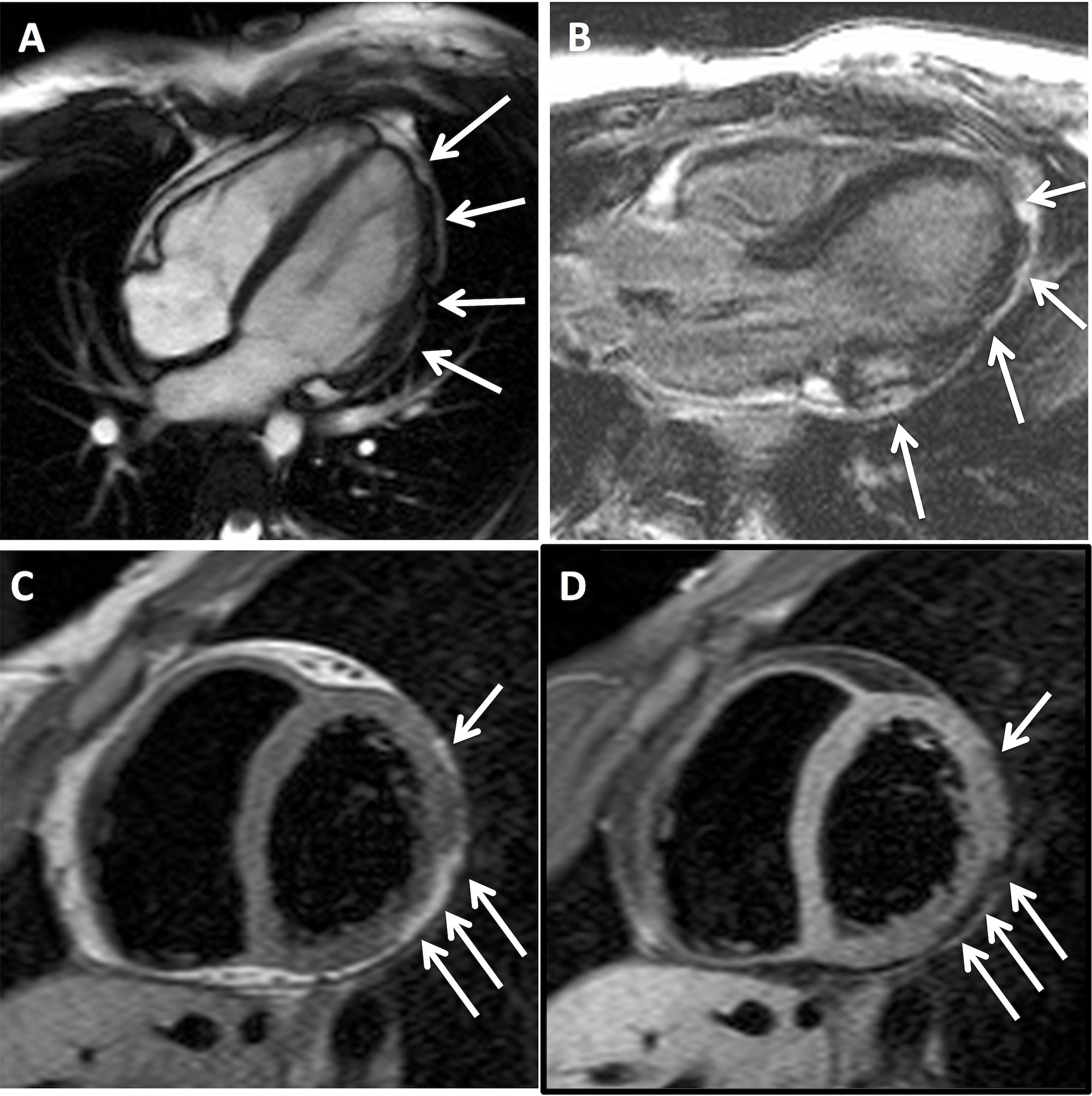
Owing to persistent symptoms, she was referred for cardiac magnetic resonance imaging (CMR). This showed mildly impaired left ventricular function, with hypokinesia and thinning / irregularity of the left antero-lateral and infero-lateral ventricular wall (Fig 1A). There was evidence of patchy epicardial late gadolinium enhancement in the lateral LV wall (Fig 1B) on the post-gadolinium sequence. No right ventricular abnormality was demonstrated. A provisional diagnosis of chronic myocarditis was made but the absence of a prolonged period of chest pain in her clinical history was felt to be unusual and an interval CMR was performed one year later. This showed nodular signal abnormality in the left ventricular lateral wall which was hyper-intense to myocardium and iso-intense to fat on T1-weighted sequences (Fig 1C) and the exhibited homogenous signal drop out with fat suppression techniques (Fig 1D), implying focal fibro-fatty infiltration.

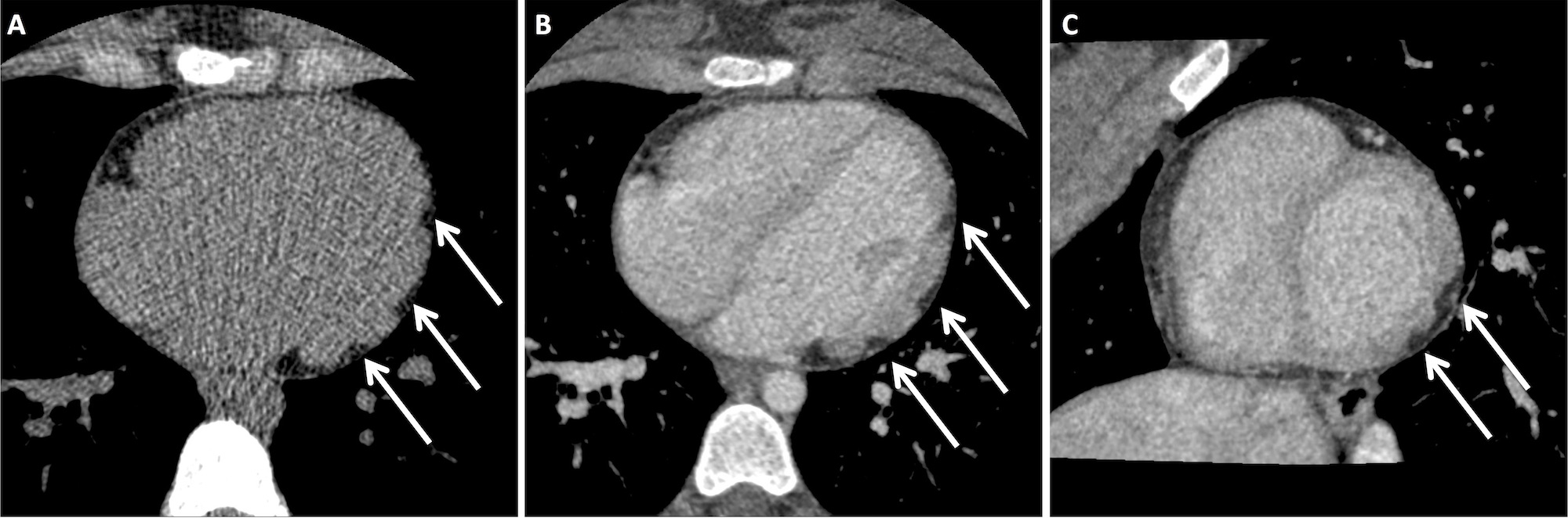
She went on to have a cardiac CT, performed with unenhanced and delayed phase imaging, to characterize the apparent nodular soft tissue seen on CMR. This confirmed abnormal lobulation of the left ventricular wall.  There were multiple areas of focal epicardial thinning.  The thin walled sections were invaginated by epicardial fat and blood vessels (Fig 2). The right ventricle was normal.

A diagnosis of arrythmogenic left ventricular cardiomyopathy (ALVC) was made. The patient was treated with bisoprolol to control palpitations and has remained well with no decline in left ventricular function.  First-degree relatives have been screened with no other affected family members identified.

Figure legends

Figure 1

A) 4-chamber steady state free precession image demonstrating irregularities of  left ventricular antero-lateral left ventricular wall (white arrows).

B) 3-chamber late gadolinium enhancement magnitude image demonstrating foci of epicardial enhancement in the infero-lateral left ventricular wall (white arrows).

C) Mid-ventricular short-axis T1-weighted image demonstrating epicardial foci of signal hyper-intense to myocardium and iso-intense to fat (white arrows).

D) Mid-ventricular short-axis T1-weighted fat-saturated image demonstrating homogenous signal drop out in the epicardial foci demonstrating high T1-weighted images in Figure 1C consistent with foci of epicardial fat.


Figure 2

A) Axial unenhanced ECG-gated cardiac CT image demonstrating foci of fat attenuation in the left ventricular lateral wall (white arrows).

B) Axial delayed ECG-gated cardiac CT image demonstrating foci of fat attenuation discrete to the enhancing myocardium in the left ventricular lateral wall (white arrows).

C) Reconstructed left ventricular short-axis delayed ECG-gated cardiac CT image demonstrating foci of fat attenuation discrete to the enhancing myocardium in the left ventricular lateral wall (white arrows).


Discussion

ALVC is an increasingly recognised condition, characterised by fibro-fatty replacement in the left ventricle wall.  It is closely related to the more widely recognised arrythmogenic right ventricular cardiomyopathy, where the right ventricle is affected. The two disease patterns frequently co-exist within the same family and genes implicated in ARVC have also been identified in patients with ALVC.

Arrythmogenic cardiomyopathies are distinct from dilated cardiomyopathy (DCM) by predominately resulting in arrhythmia, rather than ventricular dysfunction. This is an important clinical diagnosis to be aware of as the major complication is sudden cardiac death due to malignant arrhythmia. Clinical heart failure is rarely seen.


Multiple-choice questions

Question 1:

Which of these is a CMR feature of myocarditis?

A) Focal, non-ischaemic lesion demonstrating late gadolinium enhancement

B) T2 signal decrease in myocardium

C) Decreased global myocardial early gadolinium enhancement ratio.

D) Intramyocardial fat


Answer:

A) Focal, non-ischaemic lesion demonstrating late gadolinium enhancement

The Lake Louise CMR criteria1 are used to diagnose myocarditis. If ≥ 2 of the following features are present, in the correct clinical context, then a diagnosis of myocarditis should be considered:

1) the presence of at least one, focal, non-ischaemic lesion demonstrating late gadolinium enhancement.

2) regional / global myocardial signal increase on T2-weighted images.

3) increased global myocardial early gadolinium enhancement ratio (EGEr).


Question 2:

Mutations in which of these genes is most frequently associated with arrythmogenic cardiomyopathy?

A) Collagen IV

B) Desmoplakin

C) Laminin

D) Plectin


Answer:

B) Desmoplakin

When a diagnosis of arrythmogenic cardiomyopathy is made, it is important to perform familial screening. This may include a genetic screen for genotype mutations in genes such as desmoplakin2 which may be implicated in the clinical phenotype in the proband.


Question 3:

What pattern of late gadolinium enhancement would be most consistent with a diagnosis of idiopathic dilated cardiomyopathy?

A) Epicardial

B) Mid-wall

C) Subendocardial

D) Transmural


Answer:

B) Mid-wall

Mid-wall LGE is associated with idiopathic dilated cardiomyopathy. However, mid-wall fibrosis is only seen in approximately 30% of individuals with idiopathic DCM3. Epicardial LGE is described in ARVC and ALVC.  Subendocardial and transmural LGE are associated with ischaemic heart disease4.


Question 4:

Which of the following criteria was not part of the Modified Task Force Criteria for the diagnosis of arrhythmogenic right ventricular cardiomyopathy / dysplasia?

A ) Arrhythmias

B) Conduction / repolarization abnormalities

C) Endomyocardial biopsy

D) Family history

E) Global or region dysfunction


Answer:

C) Endoymocardial biopsy

For more information see the proposed modification of the Task Force Criteria5.


References

1 Friedrich MG, et al. Cardiovascular magnetic resonance in myocarditis: a JACC white paper. J Am Coll Cardiol. 2009; 53: 1475-87

2 Sen-Chowdhry S, et al. Left-dominant arrhythmogenic cardiomyopathy: an under-recognized clinical entity. J Am Coll Cardiol 2008; 52: 2176-87.

3 Assomull RG, et al. Cardiovascular magnetic resonance, fibrosis, and prognosis in dilated cardiomyopathy. J Am Coll Cardiol. 2006; 48: 1977-85

4 Mahrholdt H, et al. Delayed enhancement cardiovascular magnetic resonance assessment of non-ischaemic cardiomyopathies. Eur Heart J. 2005; 26: 1461-74

5 Marcus FI, et al. Diagnosis of arrhythmogenic right ventricular cardiomyopathy/dysplasia: proposed modification of the Task Force Criteria. Eur Heart J. 2010: 31: 806-14

Image of the month – July 2017 – A rare case of Vieussens’ arterial ring with multiple aneurysms and a coronary-pulmonary fistula

Submitted by Dr Carina Brolund-Napier (CT2 Core Medical Trainee), Dr Amit Parekh (Radiology SPR), Dr Rajiv Singh (Consultant Radiologist,), Dr Garrett McGann (Consultant Radiologist). Gloucestershire Hospitals NHS Foundation Trust, UK.

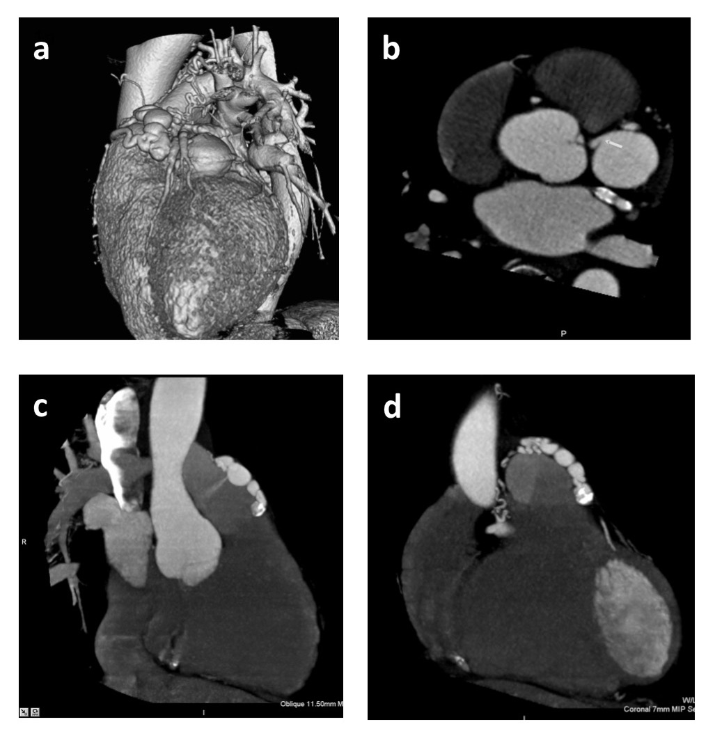
A 65 year old man presented with atypical chest discomfort and dyspnoea on a background of hypertension, hypercholesterolaemia and a positive family history of coronary artery disease. Coronary angiography showed diffuse calcific atheroma, ectatic arteries, a coronary aneurysm and a coronary-pulmonary fistula. Further imaging with computed tomography (CT) coronary angiography identified a complex fistula between a branch of the proximal left anterior descending artery feeding into a 31mm x 33mm aneurysmal segment around the right ventricular outflow tract (RVOT), in turn fistulating with the pulmonary trunk.  There was also a collateral network of dilated tortuous vessels seen anastomosing around the RVOT in keeping with a Vieussens arterial ring, a collateral pathway between the right and left coronary arteries. There was no demonstration of ischaemia on stress MRI.

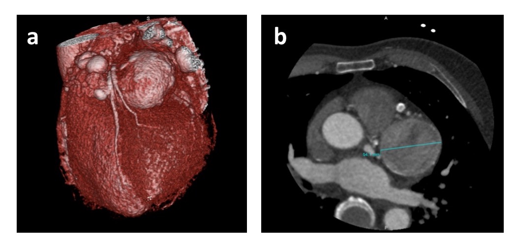
Seven years later, the patient was re-referred to cardiology with intermittent atypical chest discomfort. A repeat cardiac CT showed the aneurysm had increased in size to 51mm x 55mm with possible thrombus formation. The case was discussed at the cardiothoracic MDT where the consensus of opinion was that due to the risk of rupture this would likely require surgical repair. Further imaging with coronary angiography was recommended to ascertain if coronary intervention would be necessary at the time of surgery.

Figure 1: Imaging at first presentation

1a – Three-dimensional reconstruction CT coronary angiography. A network of dilated tortuous collateral coronary vessels with a 31mm x 33mm aneurysm lying between the circumflex and left main stem.

1b – Axial image from CT coronary angiography demonstrating the left coronary artery feeding into the coronary aneurysm.

1c – Axial image from CT coronary angiography demonstrating an aneurysmal coronary artery fistulating into the pulmonary trunk (white arrow)

1d – Axial image from CT coronary angiography demonstrating a network of dilated tortuous vessels overlying the pulmonary trunk.


Figure 2: Imaging seven years later. 

2a – Three-dimensional CT coronary angiogram image demonstrating enlargement of the main aneurysm to 51mm x 55mm.

2b – Axial image from CT coronary angiography demonstrating the aneurysm had increased in size to 51mm x 55mm. Low density is seen within the aneurysm suggesting either low flow or clot formation.


Discussion

Coronary artery fistulas (CAF) are anomalous terminations of the coronary arteries and are identified in 0.05% – 0.25% of patients who undergo coronary angiography.1 The majority of CAF are of congenital origin and adult patients are usually asymptomatic.1 Most CAF arise from the right coronary artery (50%), followed by the left coronary artery  (42%) and less commonly both the right and left coronary arteries (5%).1 CAF involving the pulmonary trunk are very rare.

Vieussens’ arterial ring (VAR) is a collateral pathway between the conus branches of the right and left coronary arteries. It is a normal anatomical variant and is rarely associated with any pathological conditions. As was the case in this patient, the presence of both a CAF into the pulmonary trunk and an aneurysm within VAR is extremely rare with very few other reported cases.2 There is one report of rupture of a 3.5cm aneurysm of VAR that was successfully treated by surgical repair.3

The management of simple CAF is controversial. In asymptomatic patients there is no urgency to close CAF and close follow-up for several years is useful. Antiplatelet and prophylactic precautions against bacterial endocarditis are recommended.1 Surgical ligation and transcatheter embolization have similar early effectiveness and patients may have an excellent prognosis following either procedure.1 However, the optimum management for large and complex CAF (as in this case) is unknown. We would therefore welcome feedback from readers about how you feel this case could be managed.


Multiple Choice Questions

  1. Which coronary vessel do the majority of symptomatic CAF arise from?
    1. LCA
    2. RCA
    3. Circumflex
    4. LAD
    5. Both RCA and LCA
  1. What is the most common clinical presentation of CAF?
    1. Dyspnoea
    2. Chest pain
    3. Sudden death
    4. Continuous heart murmur
    5. Stroke
  1. What is the chance of spontaneous closure of a CAF?
    1. 0.01%
    2. 1%-2%
    3. 10 -15%
    4. 25%
    5. 50%

References

  1. Zenooz NA. et al.  Coronary Artery Fistulas: CT findings. Radiographics 2009; 29:781-789.
  2. Lee HY, Cho SH. An Unusual Form of Coronary Artery Fistula: A Small Aneurysm of Vieussens’ Arterial Ring Communicating with the Pulmonary Artery. Korean J Thorac Cardiovasc Surg. 2014; 47:152-154
  3. Health A. Rupture of an aneurysm of Vieussens’ arterial ring presenting as acute cardiac tamponade. Clinical Radiology 2009; 64, 1129-1131


Answers1:  1 = b, 2 = d, 3 = b

Image of the Month May 2017 – Atrial septal defect and partial anomalous pulmonary venous return

Dr Lily Lei, Royal Infirmary of Edinburgh

Dr Michelle C Williams, University of Edinburgh

A 54 year old female ex-smoker presented with a ten year history of exertional breathlessness and occasional wheeze. On examination she had a soft ejection systolic murmur consistent with a pulmonary flow murmur. Electrocardiography showed right bundle branch block and echocardiography showed a superior sinus venosus atrial septal defect and a dilated right ventricle with preserved systolic function. Cardiac computed tomography demonstrated partial anomalous pulmonary venous drainage from the right upper and middle lobes. She was referred for surgery.

Atrial septal defects (ASD) are classified into three major types, namely ostium secundum, ostium primum, and sinus venosus defects.  Ostium secundum, the most common type of ASD, is a defect within the region of the fossa ovalis. Ostium primum defects are variants of atrioventricular septal defects. Sinus venosus defects are interatrial communications that usually involve the junction of the right atrium and superior vena cava 1, but less commonly can involve the venous inflow of the inferior vena cava.

The superior form of the sinus venosus ASD accounts for 5 – 10% of all ASDs 1, and is usually associated with partial anomalous pulmonary venous return (PAPVR).  PAPVR is the drainage of one or more pulmonary veins into the right atrium or systemic circulation instead of the left atrium 2. Sinus venosus ASD is associated withright-sided PAPVR, with anomalous drainage of one or more right pulmonary veins into the superior vena cava or right atrium 3.

References

1. Webb G, Gatzoulis MA. Atrial septal defects in the adult: recent progress and overview. Circulation. 2006 Oct 10;114(15):1645-53.
2. Sonavane et al. Comprehensive Imaging Review of the Superior Vena Cava. Radiographics. 2015 Nov-Dec;35(7):1873-92.
3. Porres et al. Learning from the pulmonary veins. Radiographics. 2013 Jul-Aug;33(4):999-1022.


Multiple choice questions

1. The three major types of atrial septal defects are:

A. Ostium secundum, ostium primum, ostium arteriosis

B. Ostium secundum, ostium primum, sinus venosus

C. Ostium secundum, ostium primum, sinus arteriosis

D. Ostium primum, sinus venosus, ductus arteriosis

E. Ostium primum, sinus venosus, partial anomalous pulmonary venous return


2. Which of the following is true regarding atrial septal defects:

A. Atrial septal defects are an uncommon cause of congenital heart disease

B. Downs syndrome is associated with atrioventricular septal defects

C. Sinus venosus defects are always easy to identify on transthoracic echocardiography

D. Cardiac catheterization is essential for diagnosis

E. Atrial septal defects are a common cause of cyanosis


3. Chest x-ray features of atrial septal defects may include

A. Cardiomegally

B. Enlarged central pulmonary arteries

C. Pulonary plethora

D. Small aortic knuckle

E. All of the above


Answers

1, B

2, B

3, E

Image of the Month – April 2017 – TAVI

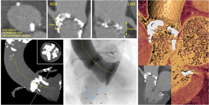
An 85-year-old woman with chronic obstruction pulmonary disease was referred to the Heart Team for consideration of transcatheter aortic valve implantation (TAVI). She had severe aortic stenosis (mean aortic valve pressure gradient 61mmHg, maximum aortic valve pressure gradient 111mmHg) with mild left ventricular systolic dysfunction. She was a life-long smoker with pulmonary function testing revealing an FEV1 1.48L. Electrocardiography demonstrated sinus rhythm with right bundle branch block and left axis deviation. Cardiac computed tomography was performed to assess suitability for TAVI.

QUESTION

What one of the following would confirm a diagnosis of severe aortic stenosis?

A. Aortic valve maximum pressure gradient 56 mmHg
B. Time to peak velocity <90 msec
C. Aortic valve calcium score 1879 AU
D. Aortic valve/LVOT velocity ratio 0.35
E. Valvulo-arterial impedance 3 mmHg/mL/m2


ANSWER C

Calcium score thresholds (men 2065 Agatston Units, women 1274 Agatston Units) differentiate moderate from severe AS [1]. Echocardiographic parameters of severe aortic stenosis (AS) include an increased aortic valve velocity (aortic valve maximum velocity 4 m/sec, maximum pressure gradient 64 mmHg (4v2), mean pressure gradient 40 mmHg). When cardiac output is reduced (i.e. LV systolic dysfunction), a velocity ratio of 4:1 (<0.25) is suggestive of severe AS. A delayed time to peak velocity (>100 msec) represents the late systolic murmur audible on auscultation that is associated with severe AS. [2] Valvulo-arterial impedance >5 mmHg/mL/m2 estimates the LV systolic load suggestive of severe AS and can be useful in patients with hypertrophied,small cavity left ventricles [3].


QUESTION

What features should be considered in this case?

A. Absolute indication for surgical bioprosthesis

B. Increased risk of left coronary artery obstruction

C. Increased risk of high-grade atrioventricular conduction block

D. Increased risk of annular rupture

E. Decreased risk of paravalvular regurgitation


ANSWER D


The correct answer is increased risk of annular rupture. This patient has a trileaflet aortic valve with severe calcification involving all three cusps. A nodule of calcification extends along the left ventricular outflow tract (LVOT) following the course of the aorto-mitral curtain. The coronary arteries arise from their normal aortic sinus with an adequate height from the annulus to the coronary ostia. There is extensive macrocalcification of the anterior (right coronary) aortic valve leaflet which may potentially obstruct the right coronary ostium during TAVI deployment. The extensive LVOT calcification beneath the left coronary artery may penetrate the myocardium and either result in acute rupture of the left ventricular free wall or slowly progress to pseudoaneurysmformation. [4]


Clinical practice guidelines would recommend transfemoralTAVI over surgical aortic valve replacement in this individual. [5] Pre-existing right bundle branch block and increased left coronary cusp calcification are independent risk factors for permanent pacemaker implantations post-TAVI. [6] The shallow sinus of valsalva height would preclude use of a self-expanding valve. [7] Extensive aortic valve calcification is associated with post-procedural prosthesis eccentricity and severity of paravalvular regurgitation [8].


QUESTION

If annular rupture were to occur, which location is most likely?

A. Intra-annular
B. Free myocardial wall
C. Below non-coronary sinus of Valsalva
D. Interventricular septum
E. Supra-annular


ANSWER A


The native annulus is severely calcified and prominent calcified shards can protrude through the annulus following balloon-expansion TAVI. Small tears or localised rupture may be ‘contained’ by the transcatheter valve preventing further haemodynamic compromise [4]. Subannular ruptures (free myocardial wall, non-coronary cusp, interventricular septum) occur when LVOT calcification disrupts the aortic-outflow tract continuity. Free myocardial wall rupture is located beneath the anterior portion of the left coronary cusp resulting in catastrophic haemorrhage. Perforation below the non-coronary sinus with calcification in the aorto-mitral continuity can generate large shunts into the left atrium. Interventricularseptum perforations may only be detected due to presence of a new conduction abnormality, however haemotoma formation and ventricular septal defects may occur. Supra-annular ruptures may develop in shallow sinuses with injuries to the sinotubular junction or coronary ostia.


Transfemoral TAVI using a 26mm Sapien 3 prosthesis was performed. Following the procedure there was only trivial paravalvular leak and no alteration to atrioventricularconduction.


References

[1] Clavel MA, Pibarot P, Messika-Zeitoun D, et al. Impact of aortic valve calcification, as measured by MDCT, on survival in patients with aortic stenosis: results of an international registry study. J Am Coll Cardiol. 2014;64:1202-1213.


[2] Kamimura D, Hans S, Suzuki T, et al. Delayed time to peak velocity is useful for detecting severe aortic stenosis. J Am Heart Assoc. 2016;5: e003907 doi: 10.1161/JAHA.116.003907.


[3] Baumgartner H, Hung J, Bermejo J, et al. Echocardiographic assessment of valve stenosis: EAE/ASE recommendations for clinical practice. Eur  J Echocardiogr. 2009;10:1-25.


[4] Pasic M, Unbehaun A, Buz S, et al. Annular rupture during transcatheter aortic valve replacement: classification, pathophysiology, diagnostics, treatment approaches, and prevention. JACC Cardiovasc Interv. 2015;8:1-9.


[5] Vandvik PO, Otto CM, Siemieniuk RA, et al. Transcatheter or surgical aortic valve replacement for patients with severe, symptomatic, aortic stenosis at low to intermediate surgical risk: a clinical practice guideline. BMJ 2016;354:i5085. Doi:10.1136/bmj.i5085.


[6] Fujita B,  Kütting M, Seiffert M, et al.  Calcium distribution patterns of the aortic valve as a risk factor for the need of permanent pacemaker implantation after transcatheteraortic valve implantation. Eur Heart J Cardiovasc Imaging. 2016;17:1385-1393.


[7] Ribeiro HB, Webb JG, Makkar RR, et al. Predictive factors, management, and clinical outcomes of coronary obstruction following transcatheter aortic valve implantation: insights from a large multicenter registry. J Am Coll Cardiol. 2013;62:1552-1562.


[8] Bekeredijan R, Bodingbauer D, Hofmann NP, et al. The extent of aortic annulus calcification is a predictor of postprocedural eccentricity and paravalvular regurgitation: a pre- and postinterventional cardiac computed tomography angiography study. J Invasive Cardiol. 2015;27:172-180.

Image of the Month – March 2017 – An unexpected finding

By Dr Katharine Garfath-Cox

University of Southampton

A 30 year old male presented to the emergency department with atypical chest pain and a mildly elevated troponin. He was referred for a CT coronary angiogram to exclude coronary artery disease.

The CTCA was acquired using a tight bolus of contrast and a region of interest in the ascending and descending aorta to enable maximal contrast density within the coronary arteries. The coronary arteries were normal. The scan demonstrated near equal contrast density within the right and left heart chambers and a significantly dilated right atrium and right ventricle.

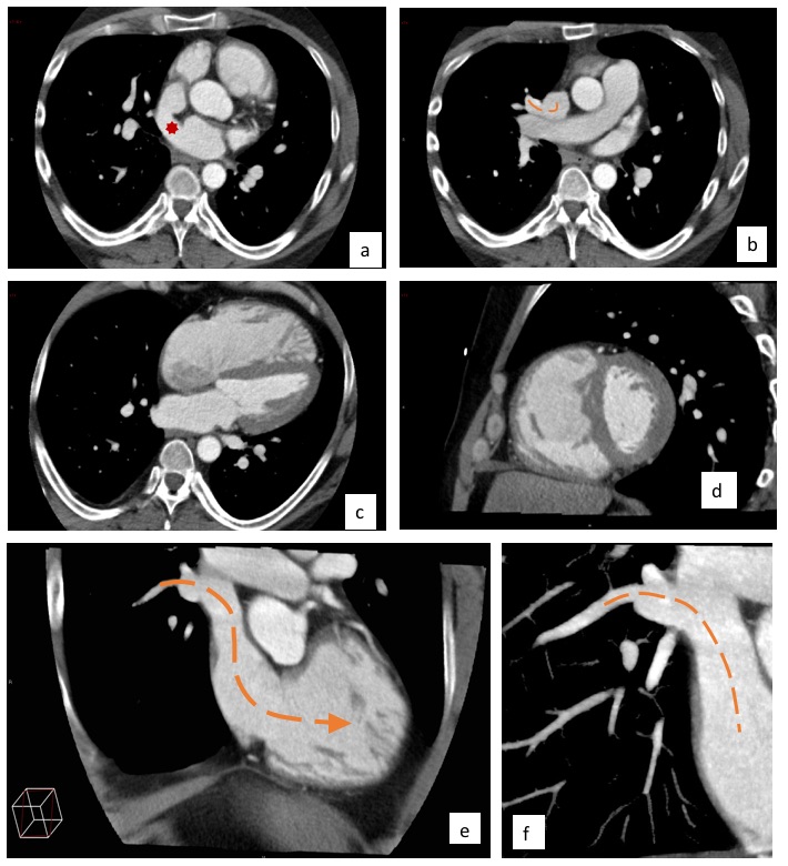
A systematic review was employed to discover the site of the presumed left to right shunt. A sinus venosus ASD (figure 1a) was present with associated anomalous drainage of the right superior pulmonary vein (figure 1b,1e,1f).

Discussion:
Atrial septal defects are the most common congenital cardiac anomaly detected in adult patients (1)(2). There are four main types, Ostium Primum, Ostium Secundum, Sinus Venosus Defect and coronary sinus type. The sinus venosus defect occurs in only approximately 5% of cases and these defects are present at the entrance of the SVC and are associated with a partial anomalous pulmonary venous connection (PAPVC).
The clinical presentation may be delayed due to low atrial pressures and even large defects can be well tolerated. The haemodynamic consequence of the defect results in a dilated right atrium and right ventricle with an increase in pulmonary vascularity. The other cardiac chambers remain normal in size.
Diagnosis is often achieved with Echo, however, with an increasing number of CT coronary angiograms being performed, review of cardiac chambers and anomalous connections is essential. The advantage of CT in this case over Echo is the concurrent identification of the site of the PAPVC, important for surgical planning.

Figure legend:
Figure1. Single beat, 16cm detector CTCA.
1a. Axial image showing a large sinus venosus ASD at the level of the entrance of the SVC into the RA (red star).
1b. Axial image of the superior pulmonary vein draining into the lateral aspect of the SVC.
1c. 4 chamber view of the dilated right atrium and right ventricle.
1d. 2 chamber view of the dilated right atrium and right ventricle.
1e. Coronal reformatted image showing the PAPVC.
1f. Cropped coronal reformat of the PAPVC.

Multiple choice questions

1. Which is the most common type of ASD?

A) Ostium primum ASD
B) Superior sinus venosus defect
C) Inferior sinus venosus defect
D) Unroofed coronary sinus type
E) Ostium Secundum ASD

2. What are the haemodynamics of an ASD?

Right atrium Right ventricle Left atrium Left ventricle  Pulmonary vasculature Aorta
A Dilated  Dilated  Unchanged  Unchanged  Increasde  Normal
B Unchanged  Dilated  Unchanged  Unchanged  Decreased  Normal
C Dilated  Dilated  Unchanged  Unchanged  Normal  Normal
D Normal  Normal  Dilated  Dilated  Increased  Enlarged
E Dilated  Dilated  Dilated  Unchanged  Increased Normal


3. This patient re-presents to the emergency department prior to intervention with breathlessness and mild cyanosis. Reversal of the shunt is identified on echocardiogram and a diagnosis of Eisenmenger physiology is made. What finding would be a feature on imaging due to the resultant pulmonary arterial hypertension?

A) Dilated pulmonary veins
B) Small central pulmonary artery
C) Mural calcification in main pulmonary arteries
D) Mural calcification in main pulmonary veins
E) Mural calcification in peripheral pulmonary arteries

4. Which of the following statements is not true regarding PAPVC?

A) Scimitar syndrome is a type of PAPVC
B) Common connections can be classified as supra-cardiac, cardiac or infra-cardiac.
C) 15% have an associated ASD
D) All pulmonary veins drain into the systemic circulation.
E) PAPVC is not always cyanotic

Images with thanks to Dr Russell Bull, Royal Bournemouth Hospital.


Answers

(1 E), (2 A, B= Tetralogy of Fallot , C=Ebstein anomaly, D=PDA), (3 C), (4 D)

Image of the Month February 2017 – A rare case of follicular thyroid carcinoma with cardiac metastasis

By: Dr Alexia Farrugia, Dr Rebecca Preston

Guy’s & St. Thomas’ NHS Trust, London

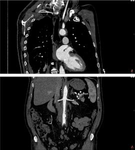
A 70 year old lady presents with shortness of breath. A chest X-ray revealed deviation of the thyroid gland to the right secondary due to a predominantly left-sided anterior mediastinal mass. CT thorax revealed a thyroid goitre. A thyroidectomy was performed and histology revealed follicular thyroid carcinoma. In view of the relatively de-differentiated tumour, radioiodine ablation was done.

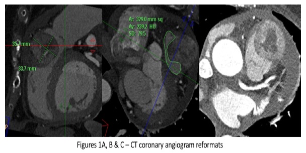
Two years later, the patient was noted on biochemical results to have a high serum calcium level and a rising thyroglobulin. FDG PET-CT was performed showing new focal increased tracer uptake posterior to the left thyroid bed and adjacent to the oesophagus. Also, there was a further focus of increased tracer uptake related to the right ventricle and pulmonary trunk. Appearances were in keeping with local recurrence within the left thyroid bed and an indeterminate new focal uptake within the right ventricle and pulmonary trunk. An urgent fine needle aspiration of the left thyroid nodule was performed and follicular thyroid carcinoma recurrence was confirmed on histology. A CT coronary angiogram revealed a soft-tissue mass anterior to and invading the pulmonary outflow tract and was seen to involve the anterior pericardium. A new pericardial effusion was noted. (Figure).

CT-guided biopsy of the anterior mediastinal mass was performed. Histology revealed round cells strongly positive for TTF-1 and thyroglobulin. Given the histological morphological features and immuno-histochemical profile of the biopsy specimen was consistent with metastatic disease from the known thyroid primary. Neck dissection was performed to excise the suspicious foci of recurrence within the thyroid bed.


Multiple choice questions

1. Which is the most common site of metastatic deposits from thyroid carcinoma?

  1. Cardiac / valvular
  2. Cervical lymph nodes
  3. Lung parenchyma
  4. Osseous
  5. Hepatic

2. Which is the commonest type of thyroid malignancy?

  1. Papillary
  2. Follicular
  3. Anaplastic
  4. Medullary
  5. Lymphoma

3. Which are the commonest tumours to metastasise to the heart?

  1. Pleural mesothelioma
  2. Melanoma
  3. Lung adenocarcinoma
  4. Breast carcinoma
  5. Squamous cell carcinoma of the head and neck

Figures 1A, B & C – CT coronary angiogram reformats. Sagittal, coronal and axial CT coronary angiogram images demonstrating a peripherally-enhancing, centrally necrotic mass within the pulmonary outflow tract, with a moderately-sized pericardial effusion.

Figures 2A & B – 18-F FDG PET-CT images. FDG PET-CT uptake is present within the right ventricular outflow tract and within the left thyroid bed consistent with a metastatic deposit and recurrence of follicular thyroid carcinoma respectively.


Images courtesy of: Guy’s & St Thomas’ NHS Trust & PET Centre at St Thomas’ Hospital, London


Answers: 1=b, 2=b, 3=a.

Image of the Month – January 2017 – Combined constrictive and restrictive pathophysiology secondary to pericardial calcification

Will Loughborough, Stephen Lyen, Mark Hamilton

University Hospitals Bristol NHS Foundation Trust

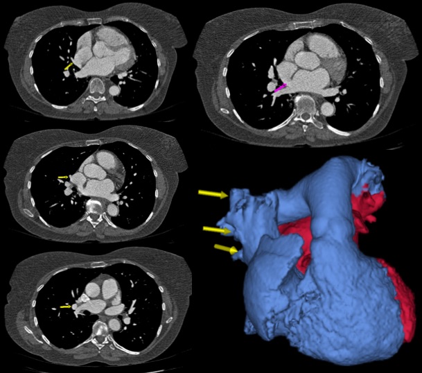
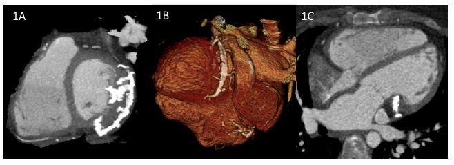
A 30 year old woman with Tetralogy of Fallot was referred for a cardiac computed tomographic examination prior to cardiac surgery. She had a Tetralogy repair in childhood involving a transannular patch. She had developed severe pulmonary valve regurgitation and was undergoing work up for an open pulmonary valve replacement which included coronary arterial assessment. A cardiac MRI demonstrated restrictive RV physiology with early diastolic septal interaction and an end diastolic A wave in the main pulmonary artery flow curve. There was unexplained reduction in LV long axis shortening both on MRI and TTE with probably pseudonormal transmitral Doppler.  A prospectively ECG gated coronary angiogram demonstrated unobstructed coronaries with normal origins. However, it revealed a thick slab of pericardial calcification extending along the posterior atrioventricular groove, with multiple prongs of calcification extending into the basal inferior and lateral left ventricular myocardium (see images) and a single prong into the RV myocardium.

On review, the extensive LV prongs corresponded to tethering of the left ventricle on dynamic imaging with consequent reduced long axis excursion. Both atria and their draining veins were dilated, implying elevated pressure (no shunt). The interatrial septum was central suggesting the pressure was equal.  The right heart findings could be explained by the RV physiology. However, these do not explain the LA findings which would seem most elegantly explained by reduced LV filling, secondary to impaired LV long axis function with secondary diastolic dysfunction. At the same time, the pericardium compressed the LV diameter a little. The conclusion was that the RV dysfunction was substantially due to the pulmonary valve disease. However the LV anatomical and physiological parameters support a diagnosis of mild systolic and diastolic dysfunction. The calcified pericardium caused pericardial constriction leading to impaired ventricular filling. The prongs of calcified pericardium invading the LV myocardium caused restriction on long axis shortening and lengthening (leading to restrictive physiology).

The patient was discussed at the adult congenital cardiac MDT where the decision was to replace the pulmonary valve alone as extensive surgical debridement of the LV pericardium was not considered attractive.

Figure 1

Figure legend

1A Short axis view. Multiple prongs of pericardial calcification extending into basal inferior and lateral left ventricular myocardium

1B Volume rendered image. Thick slab of pericardial calcification extending down the posterior atrioventricular groove

1C Four chamber long axis view. Pericardial calcification extending the basal left ventricular myocardium


Multiple choice questions

1. Treatment of Tetralogy of Fallot with a transannular patch is often associated with regurgitation though which valve?

  1. Tricuspid
  2. Pulmonary
  3. Mitral
  4. Aortic

2. Constrictive pathophysiology is usually due to abnormalities in the

  1. Myocardium
  2. Valves
  3. Coronaries
  4. Pericardium

3. Restrictive pathophysiology is usually primarily due to abnormalities in the

  1. Myocardium
  2. Pericardium


Answers

1 – B,  2 – D, 3 – A

Image of the Month – August 2016 – Platypnoea-Orthodeoxia Syndrome on perfusion scintigraphy

Submitted by Parekh A, Turner M, Gamble E, Hamilton MCK. Bristol Royal Infirmary, Upper Maudlin Street, Bristol, UK

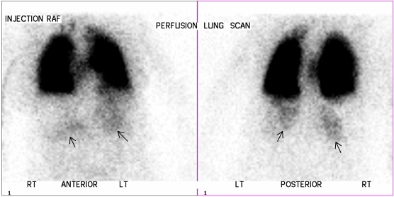
A 76 year old lady who presented with hypoxia and shortness of breath was suspected to have pulmonary embolism. She underwent perfusion scintigraphy with Technetium 99m labelled human albumin macro-aggregates (MAA). Normally, the MAA particles lodge in the pulmonary circulation and do not enter the systematic arterial circulation. In this case there was normal lung perfusion but faint tracer uptake in both kidneys (arrows) indicating a right to left shunt though a previous echocardiogram was normal. Oxygen saturations were noted to be 72% on sitting and 88% on lying. A subsequent bubble echocardiogram confirmed a right to left atrial shunt, which was significantly worse on sitting than lying supine. The patient was diagnosed with platypnoea-orthodeoxia syndrome.

Platypnoea-orthodeoxia is a rare cause of a right to left shunt within a pre-existing anatomical defect (usually an atrial septal defect (ASD) or patent foramen ovale). The shunt is more severe in the erect position[1,2]. In this case on sitting or standing the atrial septum was presumably distorted by the unfolded aortic root enlarging the defect and increasing the shunt.

The patient subsequently underwent ASD closure with marked improvement in oxygen saturations and clinical status.

References

[1] Testuz, A., Roffi, M., Müller, H., Blanche, C. and Noble, S. Platypnoea-orthodeoxia syndrome: more than just a PFO.Cardiovascular Medicine. 2014;17(7–8):228–23

[2]Cheng TO. Platypnea-orthodeoxia syndrome: Etiology, differential diagnosis, and management. Cathet Cardiovasc Interv. 1999;47:64–66.

Our Sponsors