Dr Lily Lei, Royal Infirmary of Edinburgh
Dr Michelle C Williams, University of Edinburgh
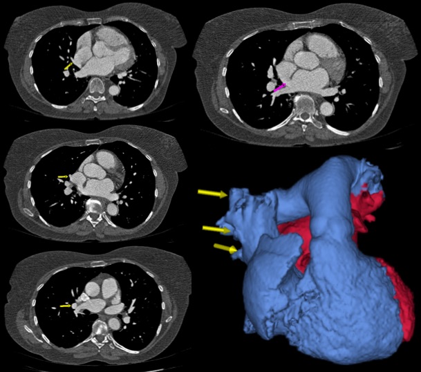
A 54 year old female ex-smoker presented with a ten year history of exertional breathlessness and occasional wheeze. On examination she had a soft ejection systolic murmur consistent with a pulmonary flow murmur. Electrocardiography showed right bundle branch block and echocardiography showed a superior sinus venosus atrial septal defect and a dilated right ventricle with preserved systolic function. Cardiac computed tomography demonstrated partial anomalous pulmonary venous drainage from the right upper and middle lobes. She was referred for surgery.
Atrial septal defects (ASD) are classified into three major types, namely ostium secundum, ostium primum, and sinus venosus defects. Ostium secundum, the most common type of ASD, is a defect within the region of the fossa ovalis. Ostium primum defects are variants of atrioventricular septal defects. Sinus venosus defects are interatrial communications that usually involve the junction of the right atrium and superior vena cava 1, but less commonly can involve the venous inflow of the inferior vena cava.
The superior form of the sinus venosus ASD accounts for 5 – 10% of all ASDs 1, and is usually associated with partial anomalous pulmonary venous return (PAPVR). PAPVR is the drainage of one or more pulmonary veins into the right atrium or systemic circulation instead of the left atrium 2. Sinus venosus ASD is associated withright-sided PAPVR, with anomalous drainage of one or more right pulmonary veins into the superior vena cava or right atrium 3.
References
Multiple choice questions
1. The three major types of atrial septal defects are:
A. Ostium secundum, ostium primum, ostium arteriosis
B. Ostium secundum, ostium primum, sinus venosus
C. Ostium secundum, ostium primum, sinus arteriosis
D. Ostium primum, sinus venosus, ductus arteriosis
E. Ostium primum, sinus venosus, partial anomalous pulmonary venous return
2. Which of the following is true regarding atrial septal defects:
A. Atrial septal defects are an uncommon cause of congenital heart disease
B. Downs syndrome is associated with atrioventricular septal defects
C. Sinus venosus defects are always easy to identify on transthoracic echocardiography
D. Cardiac catheterization is essential for diagnosis
E. Atrial septal defects are a common cause of cyanosis
3. Chest x-ray features of atrial septal defects may include
A. Cardiomegally
B. Enlarged central pulmonary arteries
C. Pulonary plethora
D. Small aortic knuckle
E. All of the above
Answers
1, B
2, B
3, E


