Submitted by Parekh A, Turner M, Gamble E, Hamilton MCK. Bristol Royal Infirmary, Upper Maudlin Street, Bristol, UK
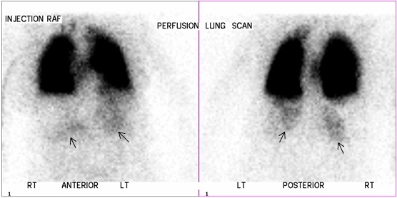
A 76 year old lady who presented with hypoxia and shortness of breath was suspected to have pulmonary embolism. She underwent perfusion scintigraphy with Technetium 99m labelled human albumin macro-aggregates (MAA). Normally, the MAA particles lodge in the pulmonary circulation and do not enter the systematic arterial circulation. In this case there was normal lung perfusion but faint tracer uptake in both kidneys (arrows) indicating a right to left shunt though a previous echocardiogram was normal. Oxygen saturations were noted to be 72% on sitting and 88% on lying. A subsequent bubble echocardiogram confirmed a right to left atrial shunt, which was significantly worse on sitting than lying supine. The patient was diagnosed with platypnoea-orthodeoxia syndrome.
Platypnoea-orthodeoxia is a rare cause of a right to left shunt within a pre-existing anatomical defect (usually an atrial septal defect (ASD) or patent foramen ovale). The shunt is more severe in the erect position[1,2]. In this case on sitting or standing the atrial septum was presumably distorted by the unfolded aortic root enlarging the defect and increasing the shunt.
The patient subsequently underwent ASD closure with marked improvement in oxygen saturations and clinical status.
References
[1] Testuz, A., Roffi, M., Müller, H., Blanche, C. and Noble, S. Platypnoea-orthodeoxia syndrome: more than just a PFO.Cardiovascular Medicine. 2014;17(7–8):228–23
[2]Cheng TO. Platypnea-orthodeoxia syndrome: Etiology, differential diagnosis, and management. Cathet Cardiovasc Interv. 1999;47:64–66.


