Image of the Month June 2020- Single Coronary Artery System or a Very Hyperdominant Left Circumflex

Single Coronary Artery System or a Very Hyperdominant Left Circumflex?


Authors and affiliations:
1. Uzma Gul
Cardiology Registrar , Russells Hall hospital
2. Robert Hugget
Consultant Cardiologist, Russells Hall hospital
3. Min Yen Wong
Consultant Radiologist, Russells Hall hospital
4. Nazia Kaushal
Consultant Radiologist, Russells Hall hospital

Case Report

A 63 year old female presented with atypical angina. Physical examination was unremarkable.
Electrocardiogram was normal and the echocardiogram revealed a structurally normal heart.
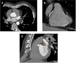
CT coronary angiography was performed and is presented below.

Images
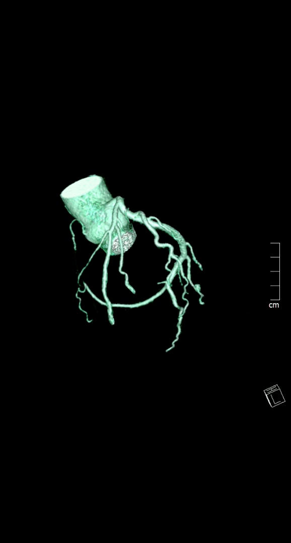
Image 1- 3D coronary tree reconstruction frontal view
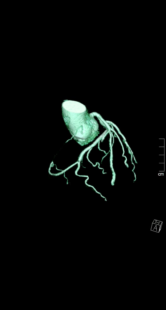
Image 2-3D coronary tree reconstruction lateral view
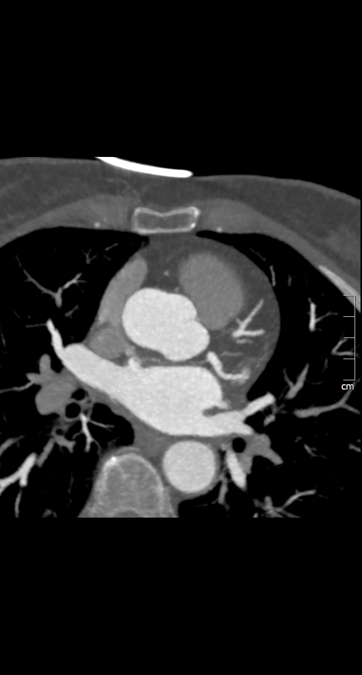
 

Image 3- Nodal artery arising from LCX
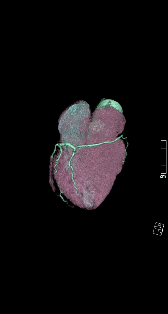
 

Image 4- 3D whole heart reconstruction

Questions & Best answers

I) What is the mechanism of ischemia in SCA?
1. Slit-like ostium & ostial ridge/ acute-angle takeoff
2. coronary spasm/ hypoplasia/ tortuous anatomy
3. Interarterial course with exercise-related narrowing
4. Intramural course with lateral compression
5. All of above

II) In Lipton classification
1. First alphabet represents the sinus of origin
2. Roman numerals represents anatomical distribution
3. Letters A,B,P,S, C represent course with reference to great vessels
4. All of above

III) Which of following congenital heart conditions are associated with single coronary artery?
1. Bicuspid aortic valve
2. Hypertrophic cardiomyopathy
3. Atrial septal defect
4. Tetrology of fallot
5. Ebstein anomaly


Answers
I) 5
II) 4
III) 1, 2 &4


Discussion

Single coronary artery (SCA) is described as an anomaly where one coronary artery originates
from a single coronary ostium from aorta and supplies the whole heart. 1 SCA is a rare anomaly
with prevalence of 0.024–0.066% in structurally normal hearts. 2 Using the Lipton classification
of SCA, our case would be best described as a type L-I SCA. 3 Type L-I is usually believed to be
benign type SCA, some malignant types are associated with sudden cardiac death. 3
However by convention, our case may also be argued as not being a single coronary artery,
since RCA had a separate ostium from aorta. Although, it did not give any of its usual branches
and ended shortly after its origin, acting as hypoplastic or atretic. Hypoplastic coronary
however, by definition, is a congenitally underdeveloped coronary with absence of
compensatory collateral vessels. 4 By contrast, in our case RCA territory was supplied by LCx
continuation into the right AV groove.
Alternatively, one may argue it as a hyper-dominant LCx with a vestigial RCA. 5 The
Hyperdominant LCx has been described in literature as supplying the apex in addition to RCA
territory. 5 None of the LAD territory was supplied by LCx in our case however.
The coronaries were free of any plaque or stenosis in our case. The patient was managed
conservatively and had an uneventful course over nine months follow-up. There are case
reports of patients presenting with angina and found to have single coronary system but no
significant atherosclerotic stenosis. 2,6 On the other hand, most case reports with
hyperdominant coronary did have an atherosclerotic stenosis explaining presentation with
angina. 5

Learning points/take home messages

1. Both single coronary artery and hyperdominant coronary are rare but can have
important implications in planning treatment.
2. SCA can present with angina or sudden death without coronary stenosis.
3. CTCA giving three dimensional anatomical data and details beyond luminography is
especially useful.

Keywords: Absent right coronary artery, Hyperdominant left coronary, Single coronary artery
anomaly.

References
1. Ayman Elbadawi, BasaratBaig, Islam Y. Elgendy. Single Coronary Artery Anomaly: A Case
Report and Review of Literature. CardiolTher. 2018 Jun; 7(1): 119–123.
2. Prashanth Panduranga, abdulla AmourRiyamia. Single coronary artery from right aortic
sinus in a very elderly patient. Journal of the Saudi Heart Association. 2016; 28(4):257-
260
3. Sinha SK, Aggarwal P, Mishra V, et al. Unusual trifurcation of a single left coronary
artery. Case Reports 2018;2018:bcr-2017-222213.
4.  Selma Kenar Tiryakioglu, Hakan Ozkan, Hakan Bahadir, et al. Left Main Coronary Artery
Hypoplasia in Elderly. Case Reports in Cardiology 2016; 2016: Article ID 4156581.
http://dx.doi.org/10.1155/2016/4156581
5. Agrawal N. Superdominant left-circumflex artery supplying significant proportion of RCA
and LAD territory. Case Reports 2015;2015:bcr2015210365.
6. Gitsios Gitsioudis,Evangelos Giannitsis, Waldemar Hosch, et al. Single coronary artery
with anomalous rising of the right coronary artery: a rare coronary anomaly diagnosed
by 256-multidetector computed tomography. Case reports in medicine. 2011; 2011:
108709. PMC3205733. doi:10.1155/2011/108709.
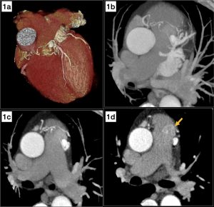
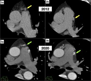
Image of the Month May 2020-The natural course of an incidentally detected coronary artery to pulmonary arterial fistula

The natural course of an incidentally detected coronary artery to pulmonary arterial fistula

Author: 

Dr Chary Duraikannu, Consultant Radiologist


Affiliations:

Countess of Chester Hospital NHS Foundation Trust, UK


Clinical history and image findings:

A 68-year-old male non-smoker with a prior history of hypercholesterolemia presents with atypical left side chest pain. Recent ECG shows sinus rhythm, with an occasional ventricular ectopic. Transthoracic echocardiography (TTE), done four months prior revealed good left ventricular function with mild mitral and tricuspid regurgitation. The patient was referred for CT coronary angiogram (CTCA) to rule out any significant coronary artery disease.

CTCA showed a bilobed aneurysmal sac (16 x 10 mm) adjoining the left side of the main pulmonary trunk, with a single large feeder from right coronary artery (likely conus branch) and multiple feeders from left anterior descending artery (Fig 1a-c). A blot of contrast density seen in the main pulmonary artery (yellow arrow) suggests the draining site (Fig 1d). The above findings were typical of a coronary artery to pulmonary artery fistula with feeders from the right and left circulation, forming an aneurysmal sac. The coronary arteries were free from atherosclerotic plaques with no significant coronary stenosis. 

Fig 1. Volume rendering (1a) and axial thick MIP (1b,c) shows an aneurysmal sac adjoining the main pulmonary trunk with feeders from the right coronary and left anterior descending arteries. Axial image (1d) shows a blot of increased contrast density in the pulmonary trunk (yellow arrow) suggesting the drainage site.

Approximately eight years prior (March 2012), the patient underwent calcium scoring, which was 0. Retrospective analysis of the images revealed a similar aneurysmal sac close to the main pulmonary trunk with adjoining feeding vessels (Fig 2,3). Despite the non-contrast scan, the findings resembled the present contrast-enhanced CT coronary angiogram and confirmed that the coronary artery fistula was indeed a long-standing finding in this patient. The cardiology team reviewed the patient and agreed that the coronary artery to pulmonary artery fistula was not the cause for patients’ current symptoms, and no further intervention was proposed.

Fig 2 & 3: Axial cardiac CT for calcium scoring (2a,b) performed in 2012 shows a similar aneurysmal sac with feeder vessels adjoining the pulmonary trunk (yellow arrows). The corresponding axial contrast enhanced CT images (3a,b) from his presentation this year is shown for comparison (green arrows).

Discussion:

Coronary artery fistula is defined as a precapillary connection between a branch of coronary artery and cardiac chamber, coronary sinus, pulmonary artery, or pulmonary vein[1]. In coronary artery fistulas, the communication can arise from the right coronary artery (50%), left coronary artery (42%) or both the coronary arterial systems (5%).[2] Though the majority of the patients are asymptomatic, some can present with chest pain, dyspnoea, and palpitations. Complications include pulmonary arterial hypertension due to the establishment of a left-to-right shunt and aneurysmal formation. Coronary artery steal phenomenon may be a theoretical risk in these patients, especially those with moderate to severe disease, for which some authors have advocated invasive FFR to assess the hemodynamic significance.[3]

Though in literature, treatment options for closure of fistula are described either by a surgical or percutaneous approach, nevertheless, there is no consensus on management strategies for coronary artery fistula.[4] In asymptomatic cases with no features of pulmonary artery hypertension or RV dysfunction, a conservative approach is wise. The case highlights the fact that even in the presence of multiple coronary artery fistulas and aneurysm formation, it would be safe to follow up in asymptomatic patients even for several years. 


Questions:

  1. Which is the most common drainage site for coronary artery fistula?
  1. Left ventricle and atrium
  2. Right ventricle and atrium
  3. Pulmonary artery
  4. Superior vena cava
  5. Coronary sinus

Answer: II. Right ventricle and atrium

The right ventricle is the most common drainage site (41%), followed by the right atrium (26%), pulmonary artery (17%). The left ventricle, left atrium, coronary sinus, and superior vena cava are rarely involved.

  1. Which of the following are complications of coronary artery fistula?
  1. Pulmonary artery hypertension
  2. Cardiac failure
  3. Aneurysm formation
  4. Myocardial ischemia
  5. All of the above

Answer: V. All of the above.

In the case of large fistulas leading to significant left to right shunt, potential complications include pulmonary artery hypertension, cardiac failure, and aneurysm formation, which can rarely rupture. There is a theoretical risk of coronary steal phenomenon leading to myocardial ischemia in these patients.

  1. Regarding the treatment of coronary artery fistulas, which of the following statements are true?
  1. In asymptomatic patients, careful periodic evaluation and follow up may be suggested
  2. Transcatheter embolization of fistula is effective
  3. Surgical ligation in coronary artery fistulas with complex anatomy
  4. Antiplatelet and Prophylactic precautions for bacterial endocarditis
  5. All of the above

Answer: V. All of the above.

There is no consensus on management strategies for coronary artery fistulas. It would be wise to follow up asymptomatic patients carefully. Both endovascular and surgical approaches are effective in the treatment of coronary artery fistula, the decision of which depends on the anatomy of the fistula.


References:

  1. Schumacher G, Roithmaier A, Lorenz HP, et al. Congenital coronary artery fistula in infancy and childhood: diagnostic and therapeutic aspects. Thorac Cardiovasc Surg 1997;45(6):287–294. 
  2. Nakamura M, Matsuoka H, Kawakami H, et al. Giant congenital coronary artery fistula to left brachial vein clearly detected by multi-detector computed tomography. Circ J 2006;70(6):796–799. 
  3. Huang Z, Liu Z, Ye S. The role of the fractional flow reserve in the coronary steal phenomenon evaluation caused by the coronary-pulmonary fistulas: case report and review of the literature. J Cardiothorac Surg. 2020 Feb 3;15(1):32. doi: 10.1186/s13019-020-1073-x. PMID: 32013986; PMCID: PMC6998067.
  4. Stout KK, Daniels CJ, Aboulhosn JA, et al. 2018 AHA/ACC guideline for the management of adults with congenital heart disease: a report of the American College of Cardiology/American Heart Association Task Force on Clinical Practice Guidelines. Circulation 2019;139:e698–800.

Image of the Month April 2020- The Consequence of a Nasty Fall

The Consequence of a Nasty Fall

Authors:

Michelle L.T. Wong and Linda Turner.

Affiliations:

Department of Radiology, Maidstone and Tunbridge Wells NHS Trust, United Kingdom.

Case Report:

A previously fit and well lady presented to the Accident & Emergency (A&E) Department after a fall down a flight of stairs. On initial assessment, she was found to be hypoxic with profound lactic acidosis.

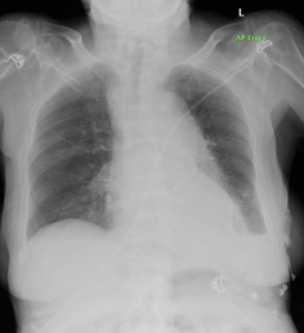
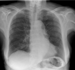
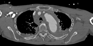
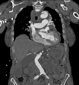
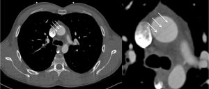
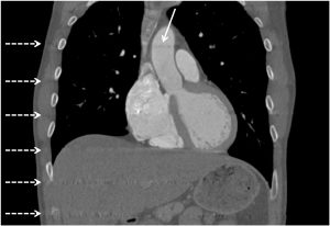
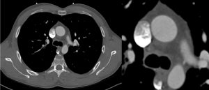
An initial chest radiograph demonstrates blunting of the left costophrenic angle with a change in the mediastinal contours (Figure 1) when compared to a previous chest radiograph performed seven years ago (Figure 2). A computed tomography (CT) aortogram was subsequently performed which demonstrates a thoracic aortic dissection flap, mediastinal haematoma and multiple rib fractures (Figures 3 and 4).

The diagnosis of traumatic acute aortic sub-adventitious tear with pseudoaneurysm formation was made. The findings were relayed urgently to the trauma and A&E team. The patient was transferred to the regional tertiary centre for further management and treatment.

Figure 1: Portable chest radiograph demonstrates blunting of the left costophrenic angle with a subtle change in the mediastinal contours when compared to a previous radiograph seven years.

Figure 2: A plain chest radiograph of the same patient that was performed seven years ago as comparison.


Figure 3: Single axial slice of the thorax from the CT aortogram that demonstrates the aortic tear with extensive mediastinal haematoma. There are associated bilateral pleural effusions.


Figure 4: Single coronal slice of the thorax (reformatted image) demonstrating the aortic tear at the level of the aortic isthmus.

Questions:

  1. Where is the commonest anatomical location for traumatic aortic injuries?
  1. Arch of the Aorta
  2. Aortic isthmus
  3. Root of aorta
  4. Descending thoracic aorta
  5. Ascending thoracic aorta

Answer: B

The aortic isthmus is the commonest location for lesions (approximately 90% of all cases of patients who have been subjected to thoracic trauma). Its relatively immobile position within the thorax due to its attachment by the ligamentum arteriosum explains the reason for this site being involved in many injuries.

    2. What is the main purpose of a chest radiograph?

  1. To detect rib fractures
  2. To detect pleural effusions
  3. To detect mediastinal haematoma
  4. To detect lung contusions
  5. To assess cardiac size

Answer: C

The main purpose is to detect a mediastinal haematoma that would indicate considerable aortic trauma. Mediastinal enlargement of more than 8cm and / or 25% of the width of the thorax is most frequently observed, but is not the most sensitive sign. The diagnosis must be suspected if there is any abnormality whatsoever of the aortic arch or opacification of the space between the aorta and the pulmonary artery.

   3. How many grades of traumatic aortic injuries are there?

  1. 5
  2. 4
  3. 3
  4. 2
  5. 1

Answer: C

Grade 1 disruption corresponds to rupture of the tunica intima and a part of the tunica media which results in a hypodense line within the aortic lumen on CT. Grade 2 disruption is a sub-adventitious rupture involving the whole of the tunica intima and media with only the tunica adventitia (a distensible and watertight structure) retaining the blood, thus forming a pseudoaneurysm. Grade 3 lesions involve ruptures of all three tunica layers of the aorta and therefore produces an extravasation of contrast agent, the aortic blood only being retained by the mediastinal fat.

References:

  1. Cullen E.L., Lantz E.J., Johnson C.M. and Young P.M. Traumatic aortic injury: CT findings, mimics and therapeutic options. Journal of Cardiothoracic Diagnosis and Therapy. 2014; 4(3): 238 – 244.
  2. Heneghan R.E., Aarabi S., Quirogi E., Gunn M.L et al. Call for a new classification system and treatment strategy in blunt aortic injury. Journal of Vascular Surgery. 2016; 64(1): 171 – 176.
  3. Yahia A.A., Bouvier A., Nedelcu C., Urdulashvili M. et al. Imaging of thoracic aortic injury. Diagnostic and Interventional Imaging. 2015; 96(1): 79 – 88.

Image of the Month March 2020- Interpolation Reconstruction Artefact On ECG-Gated CT; A Mimic Of Aortic Dissection

Authors:

Robert W. Foley, Sophie Glenn-Cox, Benjamin J. Hudson, Jonathan C. L. Rodrigues.

Affiliations:

Department of Radiology, Royal United Hospitals Bath NHS Foundation Trust, Bath, United Kingdom.

Case Report

A 36-year-old man presented to the emergency department with acute onset of severe chest pain radiating to his back. There were no high-risk exam features or a pre-existing high-risk condition. His heart rate was 40 beats per minute and regular. An urgent ECG-gated CT angiogram of the aorta was performed to exclude an acute aortic syndrome.

A retrospective spiral ECG-gated bolus-tracked acquisition (Siemens Edge, 50ml Iomeron® 350 at 5ml/sec) was performed with tube current modulation, maximum dose at 70% R-R interval and automatic best diastolic reconstruction.

The CT demonstrated a linear band of low attenuation in the ascending aorta (Figure 1).

Figure 1

The on-call general radiologist sought a specialist opinion regarding the possibility of an aortic dissection flap. However, on review of the coronal images, the linear artefacts were repeated at regular intervals along the z-axis (Figure 2).

Figure 2

The original image demonstrating the linear artefact in the ascending aorta was reconstructed at 76% of the RR interval. The cardiac radiologist suspected interpolation artefact and generated additional reconstructions after ECG editing, reconstructed at 85% of the RR interval, which significantly reduced this artefact (Figure 3).

Figure 3

Data is interpolated to generative transverse reconstructions from helically acquired CT data. It assumes a photon will experience the same attenuation if it originates from any given location or 180° from that given location. However, in reality there is slice broadening, dictated by how far the CT gantry moves during a single 180° CT beam turn. Our case highlights a rare ECG-gated artefact, which also requires acquisition to be synchronised with the R-R interval. Owing to such a slow heart rate (40 beats per minute), there was insufficient data acquired during the prolonged time between R-waves and greater distance travelled by the CT gantry per heart beat in the z-axis. The regular banding artefact resulted from interpolated data between the previous and next data points acquired by the CT during consecutive R waves [1].

The benefits of ECG-gating vastly outweigh the disadvantages of non-gated techniques when acquiring CT for suspected acute aortic syndrome. However, we present a rare case of an ECG-gating specific artefact resulting from a very low heart rate. We hope that this raises the awareness of this interpolation artefact in the setting of a very low heart rate and highlights how ECG editing can help reduce the impact and thus hopefully will mitigate against potential misinterpretation, which may have important implications for the patient.

Multiple Choice Questions

  1. Which of the following is not considered within the spectrum of Acute Aortic Syndrome?
    1. Penetrating atherosclerotic ulcer
    2. Type A aortic dissection
    3. Ruptured abdominal aortic aneurysm
    4. Type B aortic dissection
    5. Intramural haematoma
  2. According to the BSCI/BSCCT Guidelines for CT diagnosis of acute aortic syndrome [2], which of the following is not a high risk examination feature for acute aortic syndrome?
    1. Systolic blood pressure deficit
    2. Murmur of Aortic Insufficiency
    3. Focal neurological deficit
    4. Hypertension
    5. Pulse Deficit
  3. Which of the following is not a CT artefact that could be encountered when imaging in suspected acute aortic syndrome?
    1. Reverberation Artefact
    2. Pulsation artefact
    3. Interpolation artefact
    4. Breathing artefact
    5. Streak artefact
  4. In aortic dissection, which of the following does not help differentiate the true lumen from the false lumen?
    1. The beak sign
    2. The false lumen is usually smaller
    3. Intraluminal thrombosis is more commonly seen in the false lumen
    4. The intimomedial rupture sign
    5. The cobweb sign

Answers

  1. c
  2. d – Hypotension, or shock, is a high-risk examination feature for acute aortic syndrome. Hypertension is a risk factor that may lead to accelerated premature degeneration of collagen and elastin within the aorta and therefore predispose to the development of acute aortic syndrome.
  3. a – Reverberation artefact is encountered in ultrasonography. This artefact is caused by a series of delayed echoes from a strongly reflecting interface. For example, comet-tail artefact can be encountered during echocardiography in patients with prosthetic or calcified valves.
  4. b – The false lumen is usually larger than the true lumen, due to increased pressure within the dissection flap. In the beak sign, there is an acute angle between the dissection flap and the outer wall of the false lumen. The intimomedial rupture sign occurs when the entry point of a dissection flap points towards the false lumen, representing the flow of blood from true lumen to false lumen. The cobweb sign is caused by thin strands of unseparated tissue between the intima and media and can be seen within the false lumen.

References

1. Kang J-W, Do K-H, Chung J-Y, Cho HJ, Seo JB, Lim T-H (2010) Concept of minimal heart rate for each pitch value to avoid interpolation artifact when using dual-source CT: a phantom study. Int J Cardiovasc Imaging 26:103–109

2. Vardhanabhuti V, Nicol E, Morgan-Hughes G, et al (2016) Recommendations for accurate CT diagnosis of suspected acute aortic syndrome (AAS)—on behalf of the British Society of Cardiovascular Imaging (BSCI)/British Society of Cardiovascular CT (BSCCT). Br J Radiol 89:20150705

Image of the Month February 2020 – Haemoptysis and an unusual ‘lung mass’

Haemoptysis and an unusual ‘lung mass’

Tom Foster 1, Michelle C Williams 2, Peter Maclean 1

  1. Department of Radiology, Western General Hospital, Edinburgh
  2. University of Edinburgh, Edinburgh


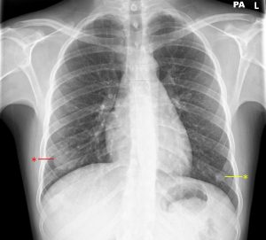
A young male patient presented to his GP after an episode of haemoptysis. He reported a 1 month history of dyspnoea, non-productive cough and intermittent fever. Initial chest X-ray (Figure 1) showed a 3.5 cm diameter rounded opacity in the right lower zone, with further patchy opacification in the left lower zone. This was initially reported as likely infective consolidation, but the clinical team’s differential diagnosis also included pulmonary embolus or malignancy.


Figure 1. 3.5 cm rounded opacity in the right lower zone (red asterisk), with further patchy opacification in the left lower zone (yellow asterisk).

Further questioning identified a history of weight loss and night sweats. Therefore a CT chest / abdomen / pelvis was performed.

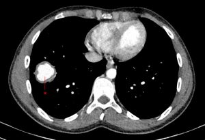
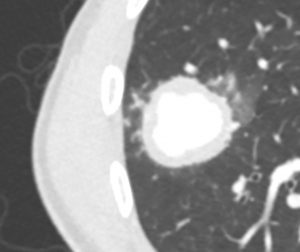
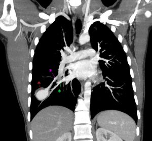
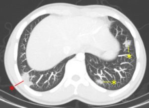
Arterial phase imaging through the lower lungs (Figure 2) showed a 3.5 cm contrast-filled lesion in keeping with a pulmonary artery aneurysm arising from a segmental branch of the right lower lobe pulmonary artery. Lung windows show some surrounding ground-glass change (Figure 3). Coronal MIP imaging (Figure 4) demonstrates a right lower lobe segmental artery pulmonary aneurysm clearly, with contrast within the lesion seen in continuity with the pulmonary trunk. A pulmonary vein is seen adjacent to the lesion, draining to the right atrium. There were a number of other areas of patchy areas of opacification located peripherally in the lungs (Figure 5). Small areas of pulmonary thrombus were seen proximal to some of these lesions. These were therefore felt to represent a mixed picture of multiple small areas of pulmonary haemorrhage, sub-segmental pulmonary emboli and focal areas of pulmonary infarction.

CT imaging of the abdomen (Figure 6) showed multiple wedge-shaped low-attenuation areas throughout both kidneys in keeping with perfusion abnormalities. There was however no other evidence of aneurysm or other significant findings in the abdomen or pelvis.

Figure 2. Axial CT slice through a contrast-filled lesion (red asterisk) in the right lower lobe on arterial-phase imaging.

Figure 3. Cropped and magnified view of the lesion on lung window showing surrounding ground-glass opacification.

Figure 4. Coronal MIP view of the aneurysm (red asterisk) of a right lower lobe segmental branch artery. The artery proximal to the aneurysm is seen superiorly (pink asterisk) and a draining pulmonary vein is seen inferiorly (green asterisk).

Figure 5. There were multiple other pulmonary abnormalities (asterisks), all located peripherally within the lungs. Some of these (red asterisk) had pulmonary thrombus proximal to them and were felt to represent pulmonary infarction.

Figure 6. An oblique coronal view through the kidneys, with multiple wedge shaped low attenuation areas bilaterally (arrowed).

The impression at this stage was that findings may represent an underlying vasculitic process such as granulomatosis with polyangiitis (previously called Wegener’s granulomatosis), Takayasu’s arteritis or Behçet’s disease. After discussion with interventional radiology and cardiothoracic surgery, the patient underwent right lower lobe basal segmentectomy.

Histopathology of the resected lung demonstrated a necrotising inflammatory process associated with areas of pulmonary infarction and focal lymphocytic vasculitis of branches of the pulmonary artery. Findings were in keeping with either Behçet’s disease or Hughes-Stovin syndrome (a rare auto-immune condition similar to Behçet’s characterised by thrombophlebitis and pulmonary/bronchial aneurysms). The patient reported oral ulcers, but no other classical findings of Behçet’s disease, therefore the final diagnosis was felt to be either atypical Behçet’s disease or Hughes-Stovin syndrome.


Discussion

Behçet’s disease is a multi-system autoimmune disease of unknown aetiology. It is more common in males and highest incidence in individuals aged 20-30, particularly those in the Middle East and Japan 1. Behçet’s Disease is likely the result of a complex interplay between various genetic and environmental factors (including certain infections and their antigens) and has been associated with HLA B51 and Factor V Leiden mutation 2.

Behçet’s disease is classically characterised by the triad of oral ulceration, genital ulceration and ocular lesions. As well as the aforementioned triad of symptoms, Behçet’s is a multi-system disorder and has the potential to involve the cardiovascular system (as in this case), the lungs, the gastrointestinal tract, genitourinary tract, the central nervous system, the skin and joints 3.

Diagnosis of Behçet’s Disease can be difficult and therefore international criteria have been developed for this purpose. The International Criteria for Behçet’s Disease (ICBD) and associated scoring is as follows, with a score of ≥4 being diagnostic for Behçet’s 4:

  • Ocular lesions – 2 points
  • Genital aphthosis – 2 points
  • Oral aphthosis – 2 points
  • Skin lesions – 1 point
  • Neurological manifestations – 1 point
  • Vascular manifestations – 1 point
  • Positive pathergy test – 1 point

The patient in this case would have scored 3 points, being positive for oral aphthous ulcers and vascular manifestations. Pathergy refers to an exaggerated reaction to skin injury from minor trauma (such as a needlestick) and this patient was noted to develop thrombophlebitis at a number of peripheral venous cannula sites but had no definite evidence of pathergy.

Treatment and prognosis is variable, with some patients’ disease resolving spontaneously, others requiring long courses of corticosteroid therapy and other’s requiring management with surgery or interventional radiology. If the disease does resolve it also has the potential to recur at a later date 5.

Hughes-Stovin syndrome is a multi-system inflammatory condition characterised by pulmonary/bronchial artery aneurysm in the setting of thrombophlebitis. Radiological findings and histopathological findings can be similar or sometimes indistinguishable from Behçet’s Disease, with the result that the term ‘incomplete Behçet’s Disease’ has been used to describe Hughes-Stovin Syndrome. Some authors postulate that Hughes-Stovin syndrome is in fact Behçet’s Disease 6 and even without the other ICBD criteria that it should be treated as such.


Questions

  1. Which human leukocyte antigen (HLA) type is associated with development of Behçet’s Disease?
    1. HLA-B27
    2. HLA-B47
    3. HLA-B51
    4. HLA-DR3
    5. HLA-DR4


2. What is the most common symptom in patients presenting with Behçet’s Disease

    1. Genital aphthous ulcers
    2. Ocular manifestations (e.g. uveitis or retinal vasculitis)
    3. Oral aphthous ulcers
    4. Joint manifestations (arthralgia or arthritis)
    5. Vascular manifestations


3. Behçet’s Disease is a vasculitis that can affect large, medium and small vessels. Which of these             vasculitides is classically considered a large vessel vasculitis?

    1. Eosinophilic granulomatosis with polyangiitis
    2. Granulomatosis with polyangiitis
    3. IgA vasculitis
    4. Kawasaki disease
    5. Takayasu’s arteritis


Answers

  1. C.

HLA-B51 is associated with the development of Behçet’s Disease 2. HLA-B27 is associated with ankylosing spondylitis. HLA-B47 is associated with congenital adrenal hyperplasia due to 21-hydroxylase deficiency. HLA-DR3 is associated with Sjogren’s syndrome, autoimmune hepatitis and type 1 diabetes. HLA-DR4 is associated with type 1 diabetes and rheumatoid arthritis 7.

  1. C.

Oral aphthous ulcers are the most common symptom in patients presenting with Behçet’s Disease. When creating the International Criteria for Behçet’s Disease (ICBD), 1278 patients with Behçet’s Disease from 27 countries were identified. 98% of these had oral aphthous ulcers, 74% had genital aphthous ulcers, 55% had ocular manifestations, 51% had joint manifestations and 19% had vascular manifestations 4.

  1. E.

Takayasu’s arteritis is a large vessel vasculitis. Eosinophilic granulomatosis with polyangiitis (previously Churg-Strauss syndrome), granulomatosis with polyangiitis (previously Wegener’s granulomatosis) and IgA vasculitis (previously Henoch-Schönlein purpura) are all small vessel vasculitides. Kawasaki disease is a medium vessel vasculitis.

_______________________

References

  1. Kontogiannis V, Powell RJ. Behçet’s disease. Postgrad Med J. 2000 Oct; 76(900): 629–637.
  2. Marshall SE. Behçet’s disease. Best Pract Res Clin Rheumatol. 2004 Jun;18(3):291-311.
  3. Davatchi F, Chams-Davatchi C et al. Behçet’s disease: epidemiology, clinical manifestations, and diagnosis. Expert Rev Clin Immunol. 2017 Jan;13(1):57-65.
  4. International Team for the Revision of the International Criteria for Behçet’s Disease (ITR-ICBD).The International Criteria for Behçet’s Disease (ICBD): a collaborative study of 27 countries on the sensitivity and specificity of the new criteria. J Eur Acad Dermatol Venereol. 2014 Mar;28(3):338-47.
  5. Cheon JH, Kim WH. An update on the diagnosis, treatment, and prognosis of intestinal Behçet’s disease. Curr Opin Rheumatol. 2015 Jan;27(1):24-31.
  6. Erkan D, Yazici Y et al. Is Hughes-Stovin syndrome Behçet’s disease? Clin Exp Rheumatol. 2004 Jul-Aug;22(4 Suppl 34):S64-8.
  7. Kumar V, Robbins SL. Table 5-7. Robbins Basic Pathology (8th edition). 2007. Philadelphia: Elsevier Saunders.

Image of the Month September 2019 – What happens when a patient’s aorta falls off?

What happens when a patient’s aorta falls off?

Dr M. Spurr, Dr A. Thorpe, Mr U. Benedetto and Dr M. Hamilton, Bristol Royal Infirmary.

A 41 year old man presented to a District General Hospital after collapsing three times at home. He had been experiencing headaches, double vision, and fever in the week prior. On admission the patient had raised inflammatory markers, a raised temperature and was tachycardic. The patient had a dental abscess one week before the symptoms started.

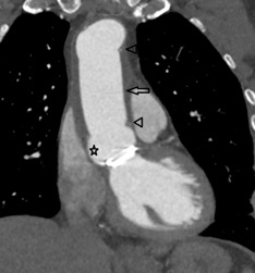
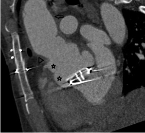
Three years previous to this admission the patient had his native bicuspid aortic valve replaced with a mechanical aortic valve and an ascending aortic replacement due to infective endocarditis (figure 1).

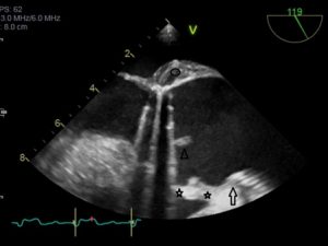
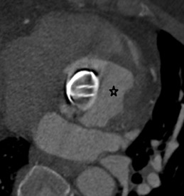
A transoesophageal echocardiogram (TOE) was performed which showed a large vegetation on the aortic valve replacement and suspicion of an aortic root abscess adjacent to the non-facing sinus (Figure 2). The anatomy was considered indeterminate on TOE. CT was initially turned down as it was thought it would not be diagnostic because of artefact from the aortic valve replacement. The patient was transferred to a tertiary hospital and a gated aortic angiogram CT was performed. The CT showed subtotal dehiscence of the aortic root from the ascending aorta with a large false aneurysm (see figures 3 and 4). The coronary insertion and proximal suture line of the ascending aortic replacement allowed the correct interpretation of the anatomy. While CT is inferior to TOE for showing vegetations, it is the test of choice if there is indeterminate anatomy or incompletely imaged aortic root pathology.

Figure 1 – Coronal oblique reformat of an aortic CT (non-gated helical). It is a baseline image from 2015 showing a post aortic valve and ascending aorta replacement

Key for figure 1

Star – sinus of Valsalva. Arrow heads – pledgets marking the proximal and distal suture lines of the ascending aortic replacement. Arrow – ascending aortic replacement.

Figure 2 – Initial transoesophageal ultrasound scan demonstrating an uncertain aortic contour

Key for figure 2

Arrow – bottom end of tube graft. Stars – “2” sinuses of Valsalva. The one closest to the aortic valve proved to be a false aneurysm. Arrowhead – aortic valve vegetation. Oval – soft tissue aortic root thickening.

Figure 3 – Reconstructed short axis CT image of the aortic root showing the “pseudo” sinus of Valsalva (star) as a large irregular cavity around the AVR

Figure 4 – Sagittal oblique ECG gated CT (systole) of the aortic root and ascending aorta

Key for figure 4

Stars – “2” sinuses of Valsalva. The one closest to the aortic valve proved to be the false aneurysm.

The patient was taken to theatre for emergency revision surgery. The aortic graft was incised and the root exposed. Direct visualisation confirmed rupture of the left sinus and an abscess associated with the right and non-facing sinus.

The previously inserted aortic graft was removed. A homograft conduit was inserted into the left ventricular outflow tract and connected distally just above the origin of coronary arteries. A tissue valve prosthesis was then implanted inside the homograft conduit. To finish a new vascular graft extending up to the proximal aortic arch was inserted.

Question 1

Which of the following statements best describes the role of CT with regards to imaging of the aortic root?

  1. TOE is the modality of choice when there is indeterminate aortic root anatomy.
  2. CT is the modality of choice to visualise an aortic valve vegetation.
  3. CT has no role in imaging of the aortic root.
  4. CT is a good alternative to visualise indeterminate aortic root anatomy if TOE is unavailable.
  5. CT is the test of choice for imaging indeterminate aortic root anatomy.

Answer – 5

As demonstrated by this case, although CT is inferior to TOE for showing vegetations, it is the test of choice if there is indeterminate anatomy or incompletely imaged aortic root pathology.

Question 2

Which of the following statements is correct regarding imaging of a patient with an AVR?

  1. An AVR is an absolute contraindication for CT imaging of the aortic root.
  2. CT should be avoided when a patient has an AVR as artefact will result in nondiagnostic images.
  3. Only a tissue valve prosthesis should be imaged with CT.
  4. Useful information can be obtained on CT despite artefact for an AVR.
  5. AVRs cause phase wrap-around artefact on CT.

Answer – 4

Useful information can still be attained despite artefact from a metallic AVR. Phase wrap-around artefact is a feature of MRI.

Question 3

What is the most common causative organism in endocarditis?

  1. Staphylococcus Aureus
  2. Streptococcus Viridians
  3. Streptococcus Mutans
  4. Escheriachia Coli
  5. Streptococcus Pneumoniae

Answer – 1 

Question 4

What percentage of patients with prosthetic valves will develop endocarditis?

  • 5%
  • 1%
  • 5%
  • 19%
  • 28%

Answer – 1

Approximately 5% of patients with prosthetic valves develop endocarditis. Involvement of the mitral valve prostheses is more frequent than that of aortic valve prostheses. (2)

References

  • Murdoch DR, Corey GR, Hoen B et al.Clinical presentation, etiology, and outcome of infective endocarditis in the 21st century: the International Collaboration on Endocarditis–Prospective Cohort Study. Archives of Internal Medicine. 2009;169(5):463–473.
  • Mylonakis E, Calderwood SB. Infective endocarditis in adults. New England Journal of Medicine. 2001;345(18):1318–1330.

Image of the Month July 2019: An unexpected finding on a non-gated CT

Authors: Tom Foster 1, Michelle C Williams 2, Marialena Gregoriades 1
  1. Department of Radiology, Western General Hospital, Edinburgh
  2. University of Edinburgh, Edinburgh

Case history:

An elderly patient was admitted with a several week history of fatigue and reduced mobility. They had previously undergone tissue AVR and CABG.

On examination they were noted to have splinter haemorrhages, Osler’s nodes and elevated inflammatory markers. This raised the suspicion of infective endocarditis (IE).

However, blood cultures were all negative and trans-thoracic echo did not demonstrate any vegetations. Trans-oesophageal echo was felt not to be indicated given negative blood cultures. A non-gated contrast enhanced computed tomography (CT) scan of the chest, abdomen and pelvis was performed to investigate infection of unknown origin.

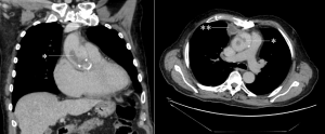
CT images identified a 45mm filling defect extending from the aortic valve into the lumen of the ascending aorta (Fig. 1). The aorta was thick walled in keep with mural oedema, suggestive of aortitis. Anteriorly in the mediastinum, anterior to the aorta and posterior to the septum there was a separate walled off, enhancing collection.

Figure 1. Coronal (left) and axial (right) CT images showing large filling defects within the ascending thoracic aorta (marked *). An enhancing collection is seen in the anterior mediastinum on the axial view (marked **).

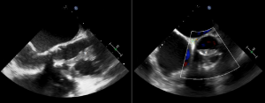
The presence of vegetations arising from the aortic valve was subsequently confirmed on trans-oesophageal echo (Fig. 2).

Figure 2. Trans-oesophageal echo showing large vegetations adjacent to the aortic valve (marked with *).

The patient was started on broad spectrum antibiotics and intravenous antifungal treatment. Further investigation with multiple blood cultures subsequently demonstrated fungal infection.

Discussion:

Infective endocarditis is infection of the inner lining of the heart, usually affecting the heart valves, with an incidence rate of approximately 1.7-6.2 cases per 100,000 patient years [1]. Despite being relatively uncommon, it is important to recognise early due to high rates of morbidity and mortality.
The most frequent aetiology is a bacterial infection, with Staphylococcus aureus and viridans group streptococci being the most common causative organisms. Other bacteria associated with endocarditis include coagulase-negative Streptococci and Enterococci, with a number of other organisms seen less frequently (such as the ‘HACEK’ group of organisms). Fungal endocarditis is less common, with Candida and Aspergillus species being the most frequent fungi identified.

Clinical presentation is variable and often linked to the underlying causative organism, with for example infective endocarditis with Staphylococcus aureus generally causes an acute presentation (<2 weeks) when compared to viridans group streptococci which are generally associated with a more subacute presentation (weeks to months). Signs and symptoms of infective endocarditis include:

  • Fever and constitutional symptoms (e.g. fatigue, malaise, etc.) – these are seen in the vast majority of patients.
  • Heart murmur – new or changing; this finding causes particular concern when associated with prosthetic valves.
  • Immunological phenomena (e.g. Osler’s nodes on the hands/feet, Roth’s spots on the retina).
  • Vascular phenomena (e.g. Janeway lesions on the hands/feet, septic emboli, splinter haemorrhages).

The modified Duke criteria (2004) can be used to diagnose infective endocarditis by assessing whether certain pathological or clinical diagnostic criteria are met. Clinical criteria involve elements of the patient history (e.g. predisposing factors), examination findings (e.g. pyrexia, Osler’s nodes and Janeway lesions), blood culture results and imaging findings. Imaging is most often in the form of echocardiography (both transthoracic and transoesophageal) but other modalities such as CT may be useful, as in this case. The European Society of Cardiology have more recently produced new modified diagnostic criteria in 2015 [2] recognise the value of cardiac CT and nuclear imaging in diagnosis.

This particular case demonstrates some of the difficulties in diagnosing infective endocarditis. Transthoracic echo in particular is limited as even large vegetations may not be identified. Prosthetic valves in particular can be challenging to image with transthoracic echo as metal artefact may obscure vegetations. It is therefore important to not be falsely reassured by negative blood cultures and a normal transthoracic echo.

While transoesophageal echo is the ‘gold standard’ imaging test for suspected infective endocarditis, this situation highlights the potential value of CT in the diagnosis.  Some patients may be too frail or not able to tolerate transoesophageal echo. Large vegetations may be identified on non-gated imaging such as in this case, but for more subtle cases gated CT may be required. Many CT scans are performed to assess sepsis of unknown origin, therefore the heart should be an important review area in such cases.

It is also important to remember fungal endocarditis as a potential source of culture-negative endocarditis. Fungal endocarditis is a rare condition but is particularly associated with immunocompromise, long term broad-spectrum antibiotic use, central venous catheters and previous cardiac surgery [3]. It is becoming increasingly common in the intensive care setting and a high index of suspicion for fungal endocarditis is important in patients in this setting who fail to respond to antibiotic therapy [2]. Embolic events are common in fungal endocarditis, more so than in bacterial endocarditis and may complicate diagnosis and subsequent management. One literature review showed that over half of cases had evidence of embolic events at initial presentation [4].


Questions:

  1. What is the most common causative organism in fungal endocarditis?
    1. Aspergillus
    2. Candida
    3. Cryptococcus
    4. Histoplasma
    5. Tricosporon
  2. What is the approximate mortality rate of fungal endocarditis?
    1. 1-2 %
    2. 10 %
    3. 25 %
    4. 30 %
    5. >50 %
  1. Which of these factors is not recognised as a predictor of poor outcome in infective endocarditis?
    1. Large vegetations
    2. Low left ventricular ejection fraction
    3. Non-HACEK Gram-negative bacilli as the causative organism
    4. Premature mitral valve closure
    5. Viridans group streptococci as the causative organism


Answers:

  1. b- Candida spp is the most common causative organism, being responsible for roughly a quarter to a half of cases of fungal endocarditis, Aspergillus spp being responsible for approximately another quarter and other fungi including Histoplasma species making up the remaining cases [5].


  1. e- Fungal endocarditis is a serious condition and fatal in approximately 50 % of cases [2]. This is for a number of reasons:
  • Difficulties in establishing a diagnosis – blood cultures are often negative. Clinical manifestations are similar to bacterial endocarditis and therefore important to consider fungal endocarditis in cases of infective endocarditis that do not respond to antibiotic therapy. Diagnosis is made post-mortem in a large number of cases [6].
  • Difficulty in treatment – treatment requires potent antifungal agents (such as amphotericin B and caspofungin) and surgical management (if patient is fit) [2]. Treatment success rate with medical therapy alone is low, and risk of recurrence is high. Specialist local microbiologist input is vital.
  • Associated comorbidities – fungal endocarditis often occurs in those with other underlying medical problems, such as those who are immunosuppressed.
  • Lack of awareness – fungal endocarditis perhaps is less well recognised than bacterial endocarditis.


  1. e- Viridans group streptococci are associated with a more subacute presentation and better prognosis. All the other answers are predictors of poor outcome [2]. Premature mitral valve closure is a feature of elevated left ventricular diastolic pressure.

References:

 [1] Beynon RP, Bahl VK, et al. Infective endocarditis. BMJ 2006; 333:334-9.

[2] Habib G, Lancellotti P, ESC Scientific Document Group et al. 2015 ESC Guidelines for the management of infective endocarditis: The Task Force for the Management of Infective Endocarditis of the European Society of Cardiology (ESC). European Heart Journal 2015; 36(44): 3075–3128.

[3] Pierrotti LC, Baddour LM. Fungal endocarditis, 1995-2000. Chest 2002; 122(1): 302–10.

[4] Kalokhe AS, Rouphael N et al. Aspergillus endocarditis: a review of the literature. Int J Infect Dis 2010; 14: 1040–1047.

[5] Ellis ME, Al-Abdely H, et al. Fungal endocarditis: evidence in the world literature, 1965-1995. Clin Infect Dis 2001; 32:50.

[6] Seelig MS, Speth CP et al. Patterns of Candida endocarditis following cardiac surgery: Importance of early diagnosis and therapy (an analysis of 91 cases). Prog Cardiovasc Dis 1974; 17(2):125-60.

Image of the Month – April 2019. Magnetic resonance angiography using feraheme (ferumoxytol)

Magnetic resonance angiography using feraheme (ferumoxytol)

Marwa Daghem, Michelle C Williams

Centre for Cardiovascular Science, University of Edinburgh, UK. 


Case

A 66 year old male with a background of hypertension and ankylosing spondylitis was admitted to the local cardiac centre with acute chest pain and a raised troponin. Electrocardiogram confirmed sinus rhythm with lateral T waves change (V4 – V6, I and aVL). He was treated for an acute coronary syndrome and subsequently had an inpatient invasive coronary angiogram (ICA). ICA confirmed the presence of three-vessel coronary artery disease with an occluded right coronary artery, moderate disease in the circumflex artery, and a diffusely diseased LAD with a moderate proximal lesion and a tight stenosis in the mid-vessel. A surgical revascularisation strategy is planned.

He agreed to participate in a research study and underwent magnetic resonance angiography using ultrasmall superparamagnetic particles of iron oxide (USPIO). Using a 3 Tesla scanner magnetic resonance coronary angiography (MRCA) was performed using 4 mg/kg ferumoxytol.

Image of the Month - April 2019. Magnetic resonance angiography using ferahaem. Figure 1

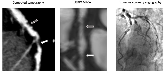
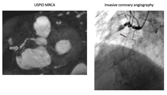
Figure 1 shows images of the left anterior descending coronary artery from computed tomography, USPIO MRCA and invasive coronary angiography. Mild non obstructive mixed plaque is seen in the proximal vessel (grey arrow). In the mid vessel there is a non-calcified plaque showing a severe stenosis (white arrow).   

Image of the Month - April 2019. Magnetic resonance angiography using ferahaem.. Figure 2

Figure 2 shows images of the right coronary artery from USPIO MRCA and invasive coronary angiograph. The right coronary artery is occluded (grey arrow). 

Image of the Month - April 2019. Magnetic resonance angiography using ferahaem. Figure 3.

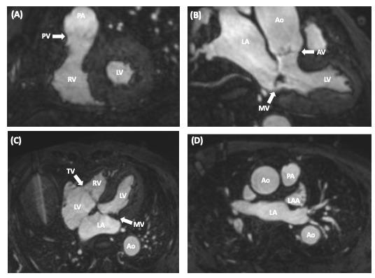
Figure 3 shows (A) short axis, (B) three chamber, (C) four chamber views of the heart showing left ventricle (LV), right ventricle (RV), right atrium (RA), left atrium (LA), left atrial appendage (LAA), pulmonary artery (PA), aorta (Ao), aortic valve (AV), pulmonary valve (PV), mitral valve (MV), and tricuspid valve (TV).


Discussion

USPIO MRCA offers an alternative to gadolinium-based MRCA. This may be useful for patients in whom gadolinium is contraindicated, such as patients with renal impairment. USPIO MRCA provides excellent identification of the presence and location of coronary arteries, down to the distal vessels and small diagonal branches. Stenosis severity can also be assessed, but with reduced accuracy compared to computed tomography coronary angiography and invasive coronary angiography.


Questions

Question 1

Based on the ECG changes and the imaging findings what vessel/segment is the likely culprit?

A. Right Coronary Artery (RCA)

B. Circumflex Artery (Cx)

C. Proximal Left Anterior Descending Artery (LAD)

D. Mid Left Anterior Descending  Artery (LAD)

Answer D.

The tight mid Left Anterior Descending Artery stenosis is the lesion that is most likely to account for this acute presentation. There is retrograde filling of the RCA suggesting this is a chronic occlusion. The LCX has only moderate disease and would not account for the ECG changes. If the proximal LAD was the culprit you would also expect to see changes in the anterior pre-precordial leads.


Question 2

Magnetic resonance coronary angiography can be performed with which of the following?

  1. Gadolinium enhanced sequences
  2. USPIO enhanced sequences
  3. Non contrast sequences
  4. All of the above

Answer D.

MRCA can be performed with gadolinium, USPIO and non-contrast sequences, although gadolinium enhanced sequences are the most widely available.


Question 3

Current indications for magnetic resonance coronary angiography include

  1. Assessment of anomalous coronary arteries
  2. First investigation for acute chest pain
  3. First investigation for stable chest pain
  4. Measurement of fractional flow reserve

Answer A.

MRCA is an option for the assessment of coronary anomalies and aneurysms, particularly in younger patients.


Question 4

Which valves have three cusps?

  1. Aortic valve
  2. Aortic and pulmonary valve
  3. Mitral valve
  4. Aortic, pulmonary and tricuspid valve

Answer D.


Acknowledgements

This research was funded by the Academy of Medical Sciences.

Image of the Month February 2019 – When a curry gives you more than heartburn..

When a curry gives you more than heartburn: A rare cause of myocardial infarction. 

Dr. S. Ahmed, Dr. A. Appaji, Dr. A. Large, Dr. S. Duckett Department of Cardiology, Royal Stoke University Hospital, Stoke-on-Trent. 

A fifty-six year old female was admitted to accident and emergency (A&E) with a witnessed collapse at home. She had a background history of recurrent urinary tract infections and an eleven pack year history of smoking. She had diarrhoea and vomiting for two days prior to admission after having a home-cooked curry and recalled feeling dizzy and nauseous with chest discomfort before passing out.

On arrival to A&E, her blood pressure was 88/62 mmHg with symptoms of feeling light-headed and clammy. On examination her jugular venous pressure was elevated, normal heart sounds and bibasal crepitations on chest auscultation. Electrocardiogram (ECG) showed normal sinus rhythm with ST segment depression in leads III, aVF and V4 to V6 with a subsequent troponin rise of > 50000. CXR showed evidence of pulmonary oedema.

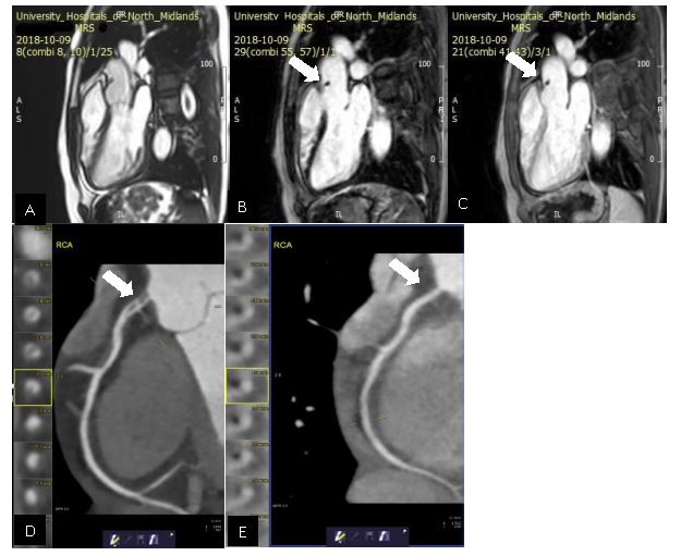
A cardiac MRI was performed which showed mild impairment of left ventricular function, a limited basal infero-septal myocardial infarction. An incidental finding of a small (5X8mm) mobile mass was seen in the proximal ascending aorta, fixed to the aortic wall superior to the ST junction and above the RCA origin which was felt to be thrombus. It was dark on the early gadolinium image (image C) and non-enhancing on the late gadolinium (image B).

CT coronary angiography was performed to further assess the coronary arteries which revealed a dissection flap (image D see arrow) causing moderate stenosis at the right coronary artery (RCA) ostium with further mild stenosis caused by a long soft tissue plaque in the proximal vessel. The images and cases were discussed with the interventional cardiology team and conservative management was felt to be the best option.

The patient was discharged with dual antiplatelet therapy for 12 months and an ACE inhibitor, statin and beta blocker. A follow up CT imaging revealed resolution of the RCA ostial dissection flap with no significant stenosis. (image E see arrow)

Image of the Month February 2019 - When a curry gives you more than heartburn...

Discussion:

Spontaneous coronary artery dissection is a rare cause of non-atherosclerotic myocardial infarction with an incidence of 0.1% to 0.4% of all acute coronary syndromes.1 It is more commonly described in young females and known to be associated with fibromuscular dysplasia, connective tissue disease and peri-partum status. Frequently identified triggers include catecholamine release mediated by physical exertion, intense emotional stress and increased abdominal pressures induced by coughing, retching or vomiting.2 Both these mechanisms increase the shearing stress on the coronary artery wall. A conservative approach towards management with standard medical therapy is usually preferred compared to invasive angiography as most cases resolve spontaneously.

References: 

1. Mortensen KH, Thuesen L, Kristensen IB, Christiansen EH. Spontaneous coronary artery dis- section: a Western Denmark Heart Registry study. Catheter Cardiovasc Interv. 2009 Nov 1;74(5):710-7.

2. Saw J, Aymong E, Sedlak T, Buller CE, Starovoytov A, Ricci D, Robinson S, Vuurmans T, Gao M, Humphries K, Mancini GB. Spontaneous coronary artery dissection: association with predisposing arteriopathies and precipitating stressors and cardiovascular outcomes. Circ Cardiovasc Interv. 2014 Oct;7(5):645-55.

Image of the Month January 2019 – Problem Solving using Cardiac CT by Modifying Technique

Problem Solving using Cardiac CT by Modifying Technique

Dr Ashley Thorpe, Dr Rajiv Singh and Dr Garrett McGann


An 80 year old presented to the emergency department after suffering syncope whilst sat in a chair with no prodromal features. Past medical history included COPD, bronchiectasis and hypothyroidism.

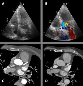
As a result, a transthoracic echocardiogram (TTE) was arranged as an outpatient which demonstrated a mildly dilated left atrium at 60mls and an echogenic structure visualised within the left atrium with a possible attachment to the atrial septum. Colour flow is seen to contour the structure.  (Figure 1A &B)

Question 1: What is the most common cardiac tumor?

  1. Lipoma
  2. Myxoma
  3. Metastases
  4. Sarcoma

Answer:

C) Metastases – Metastases are estimated to be 30 times more common than primary cardiac tumors. Common tumors which frequently metastasize to the heart include breast, melanoma and lymphoma. (1)

A CT cardiac angiogram was arranged to further investigate the ultrasound abnormality. A transoesophageal ultrasound was considered, however a CT was chosen due to its less invasive nature. A modified protocol was used to include both a prospective ECG-gated coronary artery study and a 10 second delayed phase.

During the arterial phase there was the impression of a filling defect within a mildly dilated left atrial appendage suspicious for a mass or thrombus. (Figure 1C) The critical phase in this series, the delayed 10 second phase, which showed the filling defect had disappeared suggesting the arterial phase anomaly, was caused by incomplete mixing of contrast. (Figure 1D)

Discussion:

This case highlights the importance of protocoling and making use of a delayed phase to differentiate a pseudo filling defect from true mass or thrombus. In this case the patient avoided further invasive investigation and treatment and any potential morbidity and complications they may bring. It has been shown that using a 2 phase acquisition significantly reduces pseudo filling defect detection when compared to single phase (2) and has a favourable sensitivity, specificity, positive and negative predictive values in detecting true thrombus when compared to the reference standard, trans-oesophageal echo. (3)

Question 2: What percentage of patients who have atrial fibrillation and a recent embolic event are shown to have LAA clot?

  1. 10%
  2. 15%
  3. 20%
  4. 30%
  5. 50%

Answer:

C)  ≈20%. In a study of 317 patients with AF and a recent embolic event, 21-23% were found on transoesophageal echo to have left atrial appendage clot. (5)

30-40% of strokes have no demonstrable cause. The left atrium and specifically the left atrial appendage has been studied to shed light specifically on cryptogenic stroke. Specific LAA morphologies have been shown to increase the risk of LAA thrombus and stroke. LAA function has also been assessed and poorer function has been linked to LAA thrombus and stroke.   (6)

Question 3: What are common causes of mass mimics on transthoracic echocardiography?

  1. Crista terminalis
  2. Right ventricular moderator band
  3. Atrioventricular groove fat
  4. Hiatal hernia
  5. All of the above

Answer:

E) The crista terminalis can appears as a hyperechoic structure against the right atrial wall. A thickened right ventricular moderator band can appear as a mass within the right ventricle. Lipomas of the atrioventricular groove can protrude into the atria or the ventricles and appear as an intracardiac mass. A hiatal hernia large enough can impress upon the heart and can cause a pseudomass within the left atrium. (7)


Figure 1: (A) TTE 4 chamber view demonstrating an echogenic structure within the left atrium possibly attaching to the atrial septum. (B)TTE 4 chamber view with colour flow contouring the echogenic structure within the left atrium. (C) Arterial phase CT showing a filling defect within the left atrial appendage (Arrow). (D) 10 second delay phase demonstrating resolution of the filling defect within the left atrial appendage.

Problem solving using cardiac CT


References: 

  1. Klatt EC, Heitz DR, et al. Cardiac metastases.Cancer1990; 65:1456-1459
  2. Pavitt, C& Lazoura, O, Lindsay A, et al. The Use of Cardiac CT for the Detection of Left Atrial Appendage Thrombus: A Quality Improvement Project. Heart 2014;100:A86-A87.
  3. Hur J, Kim YJ, Lee HJ, et al. Left Atrial Appendage Thrombi in Stroke Patients: Detection with Two-Phase Cardiac CT Angiography versus Transesophageal Echocardiography. Radiology 2009 251:3, 683-690.
  4. Burke A, Jeudy J, Virmani R. Cardiac tumours: an update. Heart 2008;94:117-123.
  5. Stoddard MF, Dawkins PR, Prince CR, Ammash NM. Left atrial appendage thrombus is not uncommon in patients with acute atrial fibrillation and a recent embolic event: a transoesophageal echocardiographic study. J Am Coll Cardiol 1995;25(2):452-9.
  6. Goldman ME, Pearce LA, Hart RG. Pathophysiologic correlates of thromboembolism in nonvalvular atrial fibrillation: reduced flow velocity in the left atrial appendage. J Am Soc Echocardiogr. 1999;12(12):1080-7.
  7. Malik, S. Chen, N. et al. Transthoracic echocardiography: Pitfalls and Limitations as Delineated at Cardiac CT and MR imaging. Radiographics 2017; 37:383-406
Our Sponsors