The Consequence of a Nasty Fall
Authors:
Michelle L.T. Wong and Linda Turner.
Affiliations:
Department of Radiology, Maidstone and Tunbridge Wells NHS Trust, United Kingdom.
Case Report:
A previously fit and well lady presented to the Accident & Emergency (A&E) Department after a fall down a flight of stairs. On initial assessment, she was found to be hypoxic with profound lactic acidosis.
An initial chest radiograph demonstrates blunting of the left costophrenic angle with a change in the mediastinal contours (Figure 1) when compared to a previous chest radiograph performed seven years ago (Figure 2). A computed tomography (CT) aortogram was subsequently performed which demonstrates a thoracic aortic dissection flap, mediastinal haematoma and multiple rib fractures (Figures 3 and 4).
The diagnosis of traumatic acute aortic sub-adventitious tear with pseudoaneurysm formation was made. The findings were relayed urgently to the trauma and A&E team. The patient was transferred to the regional tertiary centre for further management and treatment.
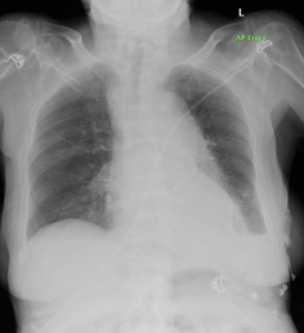
Figure 1: Portable chest radiograph demonstrates blunting of the left costophrenic angle with a subtle change in the mediastinal contours when compared to a previous radiograph seven years.
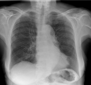
Figure 2: A plain chest radiograph of the same patient that was performed seven years ago as comparison.
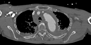
Figure 3: Single axial slice of the thorax from the CT aortogram that demonstrates the aortic tear with extensive mediastinal haematoma. There are associated bilateral pleural effusions.
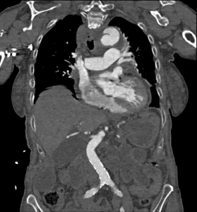
Figure 4: Single coronal slice of the thorax (reformatted image) demonstrating the aortic tear at the level of the aortic isthmus.
Questions:
- Where is the commonest anatomical location for traumatic aortic injuries?
- Arch of the Aorta
- Aortic isthmus
- Root of aorta
- Descending thoracic aorta
- Ascending thoracic aorta
Answer: B
The aortic isthmus is the commonest location for lesions (approximately 90% of all cases of patients who have been subjected to thoracic trauma). Its relatively immobile position within the thorax due to its attachment by the ligamentum arteriosum explains the reason for this site being involved in many injuries.
2. What is the main purpose of a chest radiograph?
- To detect rib fractures
- To detect pleural effusions
- To detect mediastinal haematoma
- To detect lung contusions
- To assess cardiac size
Answer: C
The main purpose is to detect a mediastinal haematoma that would indicate considerable aortic trauma. Mediastinal enlargement of more than 8cm and / or 25% of the width of the thorax is most frequently observed, but is not the most sensitive sign. The diagnosis must be suspected if there is any abnormality whatsoever of the aortic arch or opacification of the space between the aorta and the pulmonary artery.
3. How many grades of traumatic aortic injuries are there?
- 5
- 4
- 3
- 2
- 1
Answer: C
Grade 1 disruption corresponds to rupture of the tunica intima and a part of the tunica media which results in a hypodense line within the aortic lumen on CT. Grade 2 disruption is a sub-adventitious rupture involving the whole of the tunica intima and media with only the tunica adventitia (a distensible and watertight structure) retaining the blood, thus forming a pseudoaneurysm. Grade 3 lesions involve ruptures of all three tunica layers of the aorta and therefore produces an extravasation of contrast agent, the aortic blood only being retained by the mediastinal fat.
References:
- Cullen E.L., Lantz E.J., Johnson C.M. and Young P.M. Traumatic aortic injury: CT findings, mimics and therapeutic options. Journal of Cardiothoracic Diagnosis and Therapy. 2014; 4(3): 238 – 244.
- Heneghan R.E., Aarabi S., Quirogi E., Gunn M.L et al. Call for a new classification system and treatment strategy in blunt aortic injury. Journal of Vascular Surgery. 2016; 64(1): 171 – 176.
- Yahia A.A., Bouvier A., Nedelcu C., Urdulashvili M. et al. Imaging of thoracic aortic injury. Diagnostic and Interventional Imaging. 2015; 96(1): 79 – 88.


