Magnetic resonance angiography using feraheme (ferumoxytol)
Marwa Daghem, Michelle C Williams
Centre for Cardiovascular Science, University of Edinburgh, UK.
Case
A 66 year old male with a background of hypertension and ankylosing spondylitis was admitted to the local cardiac centre with acute chest pain and a raised troponin. Electrocardiogram confirmed sinus rhythm with lateral T waves change (V4 – V6, I and aVL). He was treated for an acute coronary syndrome and subsequently had an inpatient invasive coronary angiogram (ICA). ICA confirmed the presence of three-vessel coronary artery disease with an occluded right coronary artery, moderate disease in the circumflex artery, and a diffusely diseased LAD with a moderate proximal lesion and a tight stenosis in the mid-vessel. A surgical revascularisation strategy is planned.
He agreed to participate in a research study and underwent magnetic resonance angiography using ultrasmall superparamagnetic particles of iron oxide (USPIO). Using a 3 Tesla scanner magnetic resonance coronary angiography (MRCA) was performed using 4 mg/kg ferumoxytol.
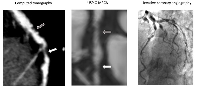
Figure 1 shows images of the left anterior descending coronary artery from computed tomography, USPIO MRCA and invasive coronary angiography. Mild non obstructive mixed plaque is seen in the proximal vessel (grey arrow). In the mid vessel there is a non-calcified plaque showing a severe stenosis (white arrow).
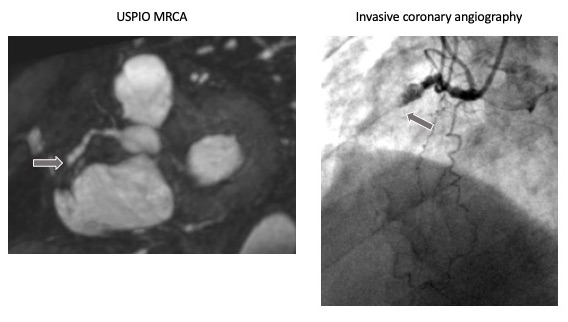
Figure 2 shows images of the right coronary artery from USPIO MRCA and invasive coronary angiograph. The right coronary artery is occluded (grey arrow).
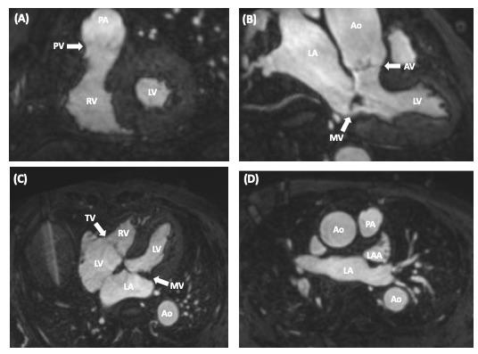
Figure 3 shows (A) short axis, (B) three chamber, (C) four chamber views of the heart showing left ventricle (LV), right ventricle (RV), right atrium (RA), left atrium (LA), left atrial appendage (LAA), pulmonary artery (PA), aorta (Ao), aortic valve (AV), pulmonary valve (PV), mitral valve (MV), and tricuspid valve (TV).
Discussion
USPIO MRCA offers an alternative to gadolinium-based MRCA. This may be useful for patients in whom gadolinium is contraindicated, such as patients with renal impairment. USPIO MRCA provides excellent identification of the presence and location of coronary arteries, down to the distal vessels and small diagonal branches. Stenosis severity can also be assessed, but with reduced accuracy compared to computed tomography coronary angiography and invasive coronary angiography.
Questions
Question 1
Based on the ECG changes and the imaging findings what vessel/segment is the likely culprit?
A. Right Coronary Artery (RCA)
B. Circumflex Artery (Cx)
C. Proximal Left Anterior Descending Artery (LAD)
D. Mid Left Anterior Descending Artery (LAD)
Answer D.
The tight mid Left Anterior Descending Artery stenosis is the lesion that is most likely to account for this acute presentation. There is retrograde filling of the RCA suggesting this is a chronic occlusion. The LCX has only moderate disease and would not account for the ECG changes. If the proximal LAD was the culprit you would also expect to see changes in the anterior pre-precordial leads.
Question 2
Magnetic resonance coronary angiography can be performed with which of the following?
- Gadolinium enhanced sequences
- USPIO enhanced sequences
- Non contrast sequences
- All of the above
Answer D.
MRCA can be performed with gadolinium, USPIO and non-contrast sequences, although gadolinium enhanced sequences are the most widely available.
Question 3
Current indications for magnetic resonance coronary angiography include
- Assessment of anomalous coronary arteries
- First investigation for acute chest pain
- First investigation for stable chest pain
- Measurement of fractional flow reserve
Answer A.
MRCA is an option for the assessment of coronary anomalies and aneurysms, particularly in younger patients.
Question 4
Which valves have three cusps?
- Aortic valve
- Aortic and pulmonary valve
- Mitral valve
- Aortic, pulmonary and tricuspid valve
Answer D.
Acknowledgements
This research was funded by the Academy of Medical Sciences.


