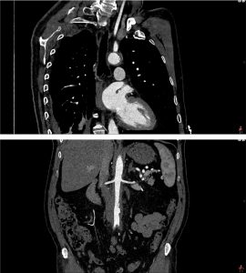
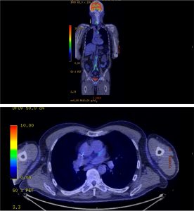
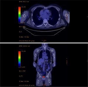
Image of the Month August 2018 – A Complicated Infarct

A Complicated Infarct

I Harries1, B Berlot1, R Ascione2, M Hamilton3, C Bucciarelli-Ducci1

1. Department of Cardiology, Bristol Heart Institute, United Kingdom

2. Department of Cardiothoracic Surgery, Bristol Heart Institute, United Kingdom

3. Department of Radiology, Bristol Royal Infirmary, United Kingdom

A 53 year old male ex-smoker with no other cardiovascular risk factors presented to a local hospital with progressive dyspnoea following an episode of chest pain that had occurred six weeks prior. ECG showed sinus rhythm with anterior Q waves. Coronary angiography demonstrated a proximal occlusion of the left anterior descending (LAD) artery and mild non-flow limiting disease in the right and left circumflex coronary arteries. The patient was commenced on appropriate medical therapy and transferred to our institution for cardiac magnetic resonance (CMR) imaging to evaluate LAD territory viability.

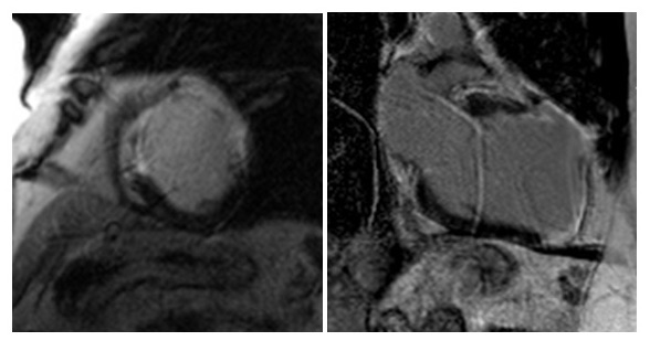
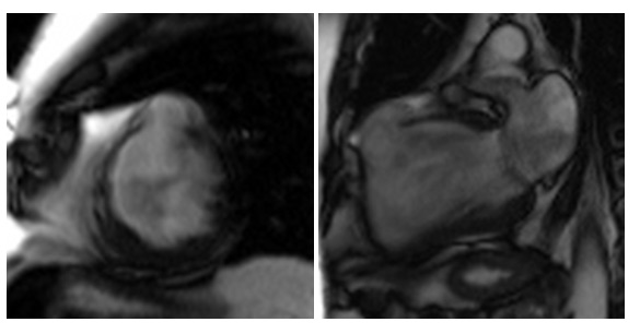
Figure 1. Mid ventricular short and 2 chamber long axis PSIR late Gadolinium enhancement sequences of the left ventricle.

  1. Is the LAD territory viable?
    1. No
    2. Yes

Answer:

  1. No. The LAD territory is not viable. There is transmural late enhancement of the mid antero-septal, mid-anterior and mid anterolateral walls on the short axis view, and of the mid and apical anterior walls and apical cap on the long axis (2 chamber) view. The likelihood of functional recovery following revascularisation is very low in segments with >75% hyperenhancement 1.

Figure 2. Mid ventricular short axis and long axis 2 chamber balanced SSFP cine stills of the left ventricle.

  1. What complication of myocardial infarction is demonstrated?
    1. Left ventricular aneurysm
    2. Ventricular septal defect
    3. Papillary muscle rupture
    4. Left ventricular pseudoaneurysm
    5. Left ventricular thrombus

Answer:

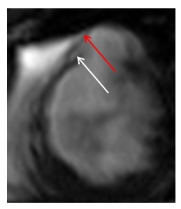
d.         Left ventricular pseudoaneurysm.  Myocardial rupture is evidenced by the abrupt discontinuation of the myocardium in the mid anterior segment (white arrow) with containment by the overlying pericardium (red arrow).


Figure 3. Zoomed mid ventricular short axis balanced SSFP cine still of the left ventricle.

A pseudoaneurysm or ‘false aneurysm’ is formed when cardiac rupture is contained by adherent pericardium or scar tissue. Unlike true aneurysms, pseudoaneurysms contain no endocardium or myocardium and are significantly more likely to rupture than true aneurysms, which rarely rupture because of the densely fibrotic tissue found in the wall of true aneurysms 2.

  1. What is the appropriate management strategy?
    1. Surgical repair
    2. Medical therapy
    3. Revascularisation of the LAD (CABG or PCI)
    4. Cardiac transplant

Answer:

  1. Surgical repair. If feasible, surgical repair is the preferred therapeutic option because conservative management of pseudoaneurysms carries a 30-45% risk of rupture and a mortality of almost 50%3. Perioperative mortality is less than 10% with greater risk for patients requiring concomitant mitral valve repair2.


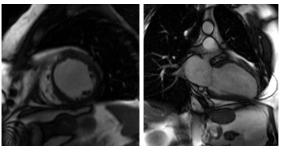
The patient successfully underwent left ventricular aneurysmectomy (Figure 4.), was established on appropriate medical therapy, had a primary prevention ICD implanted and is making a good recovery.

Figure 4. Mid ventricular short axis and long axis 2 chamber balanced SSFP cine stills of the left ventricle following left ventricular aneurysmectomy.

References

  1. Kim RJ, Wu E, Rafael A, Chen EL, Parker MA, Simonetti O, Klocke FJ, Bonow RO, Judd RM. The use of contrast-enhanced magnetic resonance imaging to identify reversible myocardial dysfunction. N Engl J Med. 2000 Nov 16;343(20):1445-53.
  2. Shapira O. (2017, Nov 27) Left ventricular aneurysm and pseudoaneurysm following acute myocardial infarction. Retrieved from https://www.ncbi.nlm.nih.gov/pmc/articles/PMC4900369/.
  3. Frances C, Romero A, Grady D. Left ventricular pseudoaneurysm. J Am Coll Cardiol. 1998 Sep;32(3):557-61.

Image of the Month July 2018 – Fluttering between imaging modalities

Dr Jamie Kitt, Dr Andrew Kelion  and Dr Elizabeth Orchard.

Department of Cardiology, Oxford Heart Centre, John Radcliffe Hospital, Oxford, United Kingdom

A 46-year-old civil servant was referred to us by a local district general hospital (DGH) in April 2018.  He had been admitted in October 2017 after developing rapid palpitations whilst in the shower. He attended his local Emergency Department and was found to be in atrial flutter with 2:1 block and a ventricular rate of 140 beats per minute. He was seen by the on-call cardiologist and started on Bisoprolol and Apixaban. He was referred to the local Electrophysiologist. An A&E bedside scan (GE V-scan) done locally was ‘reportedly normal’ and he went on to have a successful flutter (cavo-tricuspid isthmus) ablation in December 2017.

At our clinic in June 2018, he reported no further palpitations, but mentioned fatigue with reduced exercise tolerance compared to early 2017. On reflection, he felt that he had always struggled to keep up with his peers at school and university. He had no other past medical history of note, and was a non-smoker who drank only occasional alcohol. He took only Apixaban 5mg bd.

On examination, oxygen saturation was 99% on room air. Pulse rate was 66 and regular, with normal character and volume, and blood pressure was 115/60. Jugular venous pulse was normal. He had wide fixed splitting of the second heart sound, and a pan-systolic murmur loudest at the lower left sternal edge. The chest was clear.

12-lead electrocardiogram (ECG) demonstrated sinus rhythm, with partial right bundle branch block (RBBB) and a QRS duration of 100ms.

Question 1: What is the likely diagnosis at this point based on the current information?

  1. Ventricular septal defect (VSD)
  2. Atrio-ventricular septal defect (AVSD)
  3. Mitral regurgitation
  4. Atrial septal defect (ASD)
  5. Pulmonary stenosis

Answer: 4. Atrial septal defect (ASD). The combination of long-standing exercise intolerance, an atrial arrhythmia, wide fixed splitting of the second heart sound and RBBB morphology on the ECG is highly suggestive of an ASD1.

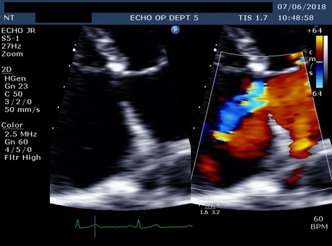
At follow up in the DGH, he had eventually had a formal trans-thoracic echo and a dilated right heart was noted as well as an atrial septal defect which prompted the referral to us. A transthoracic echocardiogram (TTE) at our clinic was reported as a primum ASD with a left to right shunt (Figure 1).  There was also moderate mitral regurgitation due to prolapse of the anterior mitral valve leaflet (AMVL) with thickening of the leaflet tips (Figure 2). The right ventricle (RV) was moderately dilated, with severe bi-atrial dilatation.

Figure 1. Trans-thoracic echo apical 4 chamber view A :  Severely enlarged left atrium  B: Severely dilated RA with evidence of RA pressure overload at end systole (arrow) C: RV dilatation (albeit in snap-shot at end-systole) D (arrow): Elongated AMVL with prolapse
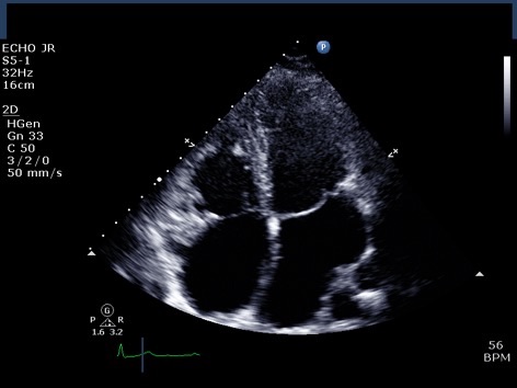
Figure 2.  A: Zoomed apical 4 chamber of the inta-atrial septum showed a defect (measured as 1.3cm) reported as a primum ASD. B: Corresponding colour flow Doppler through the defect confirming left to right shunt
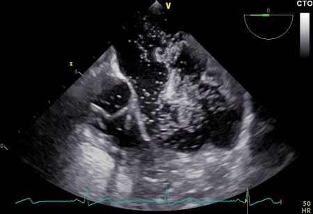
Transoesophageal echocardiography (TOE) was performed a few weeks later, and the images were reviewed at our Adult Congenital Heart Disease (ACHD) Multidisciplinary Team (MDT) Meeting. The study was felt to show a primum ASD, with an accompanying bubble study which appeared to show a left to right shunt (Figures 3A and 3B). However, the atrial defect itself was not clearly visualised, so cardiac magnetic resonance (CMR) imaging was advised to define the anatomy more precisely.

Figure 3A.  Trans-oesophageal echocardiohgram (TOE) mid-oesophageal at 71 degrees focusing on the intra-atrial septum (IAS) A: Severely enlarged right atrium. B: IAS with small primum ASD suspected but unable to clearly visualise at any angulation C: Off axis tri-leaflet aortic valve D: Partial view of a dilated RV
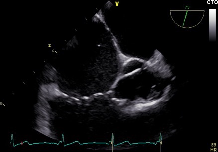
Figure 3B. Bubble study during TOE at 3rd cardiac cycle after injection into left arm (unaware of left sided SVC at this time) consistent with large left to right shunt.
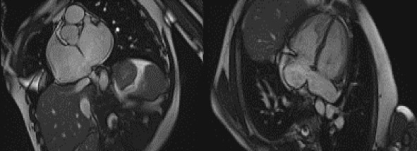
CMR was performed in July 2018, including a detailed atrial stack and a twist angiogram in addition to the standard set of sequences. The study reportedly showed a moderate secundum ASD located antero-inferiorly (Figure 4A and 4B), resulting in a 2:1 left to right shunt. A comment was made that no primum ASD was seen. There was a mildly dilated RV when indexed to his body surface area (235mls) with normal RV function, normal LV function, and mild mitral regurgitation. Normal pulmonary venous (PV) drainage was noted, but a persistent left-sided superior vena cava (SVC) was seen on the twist angiogram though it was unclear whether this was draining into the right atrium (RA).


Figure 4. Cardiac MRI performed using Siemens 1.5T magnet. 4A Atrial stack A:Severely dilated left atrium. B: Severely dilated right atrium. C (arrow): Atrial Septal defect visualised reported as moderate sized secundum atrial septal defect (ASD) measuring (21x17mm) which is located very antero-inferiorly. Flows subsequently  confirmed Qp:Qs 2:1. 4B Horizontal long axis (HLA) view also showing significant atrial septal defect (arrow) and D: moderately dilated RV
Question 2: What is the commonest variation in the thoracic venous system?

  1. Anomalous drainage of right sided superior vena cava (SVC) to the left atrium (LA)
  2. Persistent left-sided SVC (bilateral SVCs aka SVC duplication)
  3. Isolated left sided SVC

Answer: 2. Persistent left-sided SVC, is the most common congenital venous anomaly in the chest.

It accounts for 10% of thoracic vein anomalies and is present in 0.3-0.5% of the general population2. It is only seen in isolation in 10% of cases since in the vast majority of cases (82-90%) a normal (but small) right-sided SVC is also present, and a persistent bridging vein (left brachiocephalic vein) is seen in 25-35% of cases.

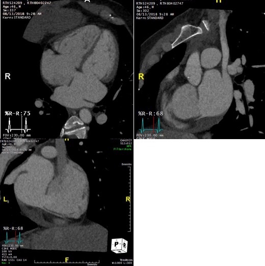
On further discussion at the ACHD MDT Meeting in August 2018, there was still no agreement on the exact anatomy despite TTE, TOE with a bubble study, and CMR. He was duly referred for a gated cardiac X-ray computed tomographic (CT) study, which resolved the confusion. CT demonstrated a right-sided SVC and inferior vena cava (IVC) draining normally into the RA. The persistent left-sided SVC (figure 5B in Powerpoint) drained into a completely de-roofed coronary sinus and LA (Figure 5), creating a large effective “ASD”. An additional small secundum ASD was also evident. PV drainage was normal. The right ventricle and both atria were dilated. The coronary arteries were normal.


Figure 5 . Gated Cardiac CT which was performed on a GE Revolution with 16cm detector, using Omnipaque 350 50ml at 5ml/s, saline 50ml at 5ml/s. No metoprolol or GTN was administered. Prospective gating at 70-80% as well as 0-100% at 20% current. 100kVp. Heart rate 64bpm. DLP 223mGy.cm.

5A: A: Deroofed coronary sinus creating large ‘ASD’ as well as a B: Secundum ASD and 5B shows C: Left sided SVC draining into De-roofed coronary sinus and Left atrium (LA).
 

Question 3: What is the commonest associated congenital lesion seen with a left sided SVC

  1. Atrial septal defect (ASD)
  2. Ventricular septal defect (VSD)
  3. Coarctation of the aorta
  4. Pulmonary stenosis
  5. Anomalous pulmonary venous return

Answer: 1. ASD. Congenital heart defects are present in 4.4% of patients with left-sided SVC3. ASDs are the most common by far but it can also occur with a single atrium, VSDs, Tetralogy of Fallot, coarctation of the aorta, anomalous pulmonary venous drainage and pulmonary stenosis. These should be thus searched for if a left sided SVC is present. There are a number of possible drainage sites. Left-sided SVCs are often functionally insignificant with 92% draining into coronary sinus since venous return from the head, neck and upper limbs is delivered to the right atrium 3. In the other 8%, although drainage into the LA results in a right to left shunt, it is usually not large enough to cause cyanosis or symptoms 3but, in this case the coronary sinus ASD and second secundum ASD resulted in a large left to right shunt.

Discussion:

This case highlights the importance of comprehensive cardiological assessment of patients with new onset atrial arrhythmias, including a detailed TTE, particularly in young adults, before jumping to treat the rhythm disorder. It also highlights the importance of searching for other lesions once one congenital lesion has been identified. Moreover, it emphasises the strengths and limitations of the various imaging modalities used in assessment of complex congenital heart disease. Echocardiography and CMR provide excellent functional information. However, when the anatomy is uncertain, gated cardiac CT offers the unparalleled ability to perform extensive reorientation of a true three-dimensional dataset with high spatial resolution. There is no requirement for images to have been acquired in precisely the right plane, as for echo and CMR. Modern CT scanners such as the GE REVOLUTION also allow easy dose modulation across the entire cardiac cycle, so that even LV and RV systolic function can be calculated to complement the anatomical assessment.

References:

  1. S Martin, E Shapiro and M Mukherjee. Atrial Septal Defects – Clinical Manifestations, Echo Assessment, and Intervention. Clin Med Insights Cardiol. 2014; 8 (Suppl 1): 93–98. PMCID: PMC4373719
  2. Padhani AR, Hale HL. Mediastinal venous anomalies: potential pitfalls in cancer diagnosis. Br J Radiol. 1998; 71 (847): 792-8. Br J Radiol
  3. Pretorius PM, Gleeson FV. Case 74: right-sided superior vena cava draining into left atrium in a patient with persistent left-sided superior vena

Image of the Month – June 2018 – An unusual cause of extensive bilateral lower limb oedema

An unusual cause of extensive bilateral lower limb oedema

Rachael Forsythe and Orwa Falah, Edinburgh

An 84 year old male presented with sudden onset massive bilateral lower limb swelling and a 4 day history of vague abdominal pain and swelling.  He had no previous similar episodes; otherwise fit and well.  Still working as a farmer. Treated for hypertension (bisoprolol), atrial fibrillation (recently commenced on apixaban) but no other relevant past medical history.  No personal or family history of thrombophilia.  He was haemodynamically stable on admission. Relevant bloods: Hb 118, platelets 160, creatinine 169, urea 16.6, eGFR 33.

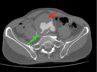
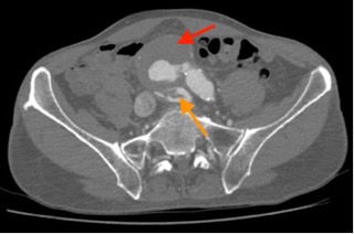
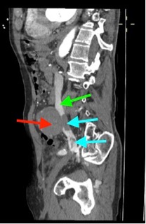
Urgent CTA was performed (images 1-3).

Image 1: Large AAA (red arrow) rupturing into IVC (green arrow) via aorto-caval fistula.

Image 2: Contrast from the AAA (red arrow) is seen within the inferior vena cava and iliac veins (orange arrow)

Image 3: Contrast from the AAA (red arrow) is seen within the inferior vena cava (green arrow), which also contains thrombus (blue arrows)

Question 1:

What are the diagnoses?

Question 2:

What effect does acute renal failure have on apixaban clearance?

  1. a.no effect – apixaban does not undergo renal excretion and would therefore be cleared within 48 hours
  2. b.increases the clearance time; dose should be reduced if serum creatinine >133micromol/litre or age >80 or weight <61kg
  3. c.increases the clearance time by an extra 72 hours but only when the eGFR is below 30

           Question 3:

 Why might a patient with aorto-caval fistula present with breathlessness and syncope?

  1. a)aorto-caval fistula can cause a left-to-right shunt resulting in high output congestive cardiac failure
  2. b)breathlessness and syncope is a manifestation of haemorrhagic shock
  3. c)they may develop acute aortic stenosis due to elevated right ventricular systolic pressure

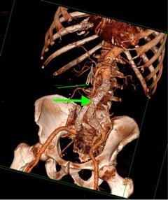
The optimal approach to this would be urgent open repair, however in the context of acute renal failure, apixaban assay confirmed that clearance was markedly reduced. It was therefore not felt safe to proceed with open repair in the acute period due to the risk of being unable to achieve haemostasis.  The patient therefore had a trans-jugular inferior vena caval filter inserted followed by standard EVAR the following day (image 4).

Image 4: 3D reconstruction following EVAR (thick green arrow), with IVC filter visible (thin green arrow)

Despite successful EVAR deployment, the fistula remained patent with a type 2 endoleak due to patent lumbar arteries.  However, renal function rapidly returned to normal and the patient experienced rapid symptomatic relief.  He was discharged home on warfarin with plans for early follow up imaging.

Answers:

  1. Ruptured abdominal aortic aneurysm with large aorto-caval fistula and concurrent bilateral ilio-femoral DVTs extending into the IVC
  2. B
  3. A

Image of the Month May 2018 – Absence doesn’t make the heart grow fonder

Absence doesn’t make the heart grow fonder:  an interesting case of partial pericardium.

Dr Tom Chance1, Dr Laura Duerden1, Dr Jonathan Rodrigues2, Dr Stephen Lyen1, Dr M Hamilton1, Dr N Manghat1.

Affiliations:

1 Department of Radiology, Bristol Royal Infirmary, United Kingdom

2 Department of Cardiothoracic Imaging, Toronto General Hospital, Toronto


Image focus:

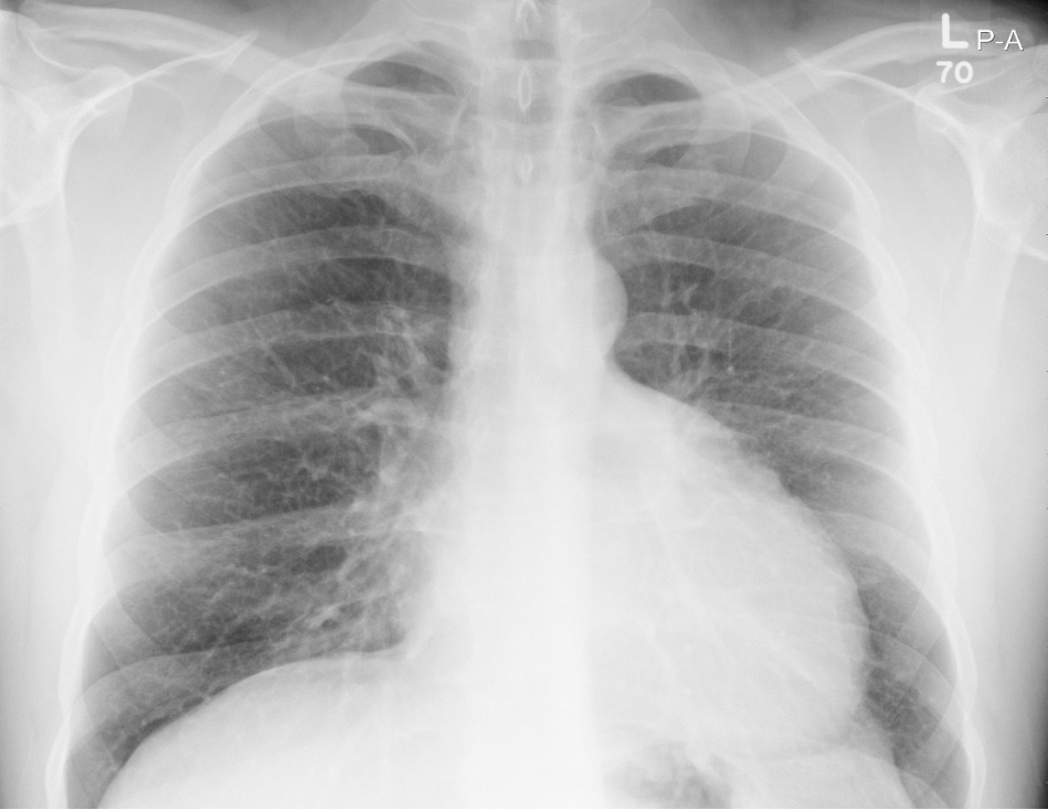
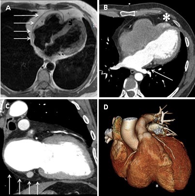
A 45 year old man presented with atrial fibrillation.  A chest radiograph showed a slightly lobulated contour of the superior left heart border (see figure 1). Transthoracic echocardiography (performed at an outside institution) showed possible right ventricular (RV) mass.  Cardiovascular magnetic resonance was performed to clarify the echocardiographic appearances. It demonstrated abrupt pericardial discontinuity on axial HASTE sequence, with the right-sided pericardial edge causing focal, extrinsic RV compression (Fig 2A). Steady state free precession cine images demonstrated an impaired RV ejection fraction of 34%.  The RV indexed end diastolic volume was 77ml/m2 (normal age-matched range 67-111ml/m2). RV volumes quantified on short axis slices. Left ventricular (LV) morphology and function were normal (LV ejection fraction 45%). Cardiac CT was performed to provide higher spatial resolution detail of the pericardial abnormality. This was performed with retrospective gating with 20% mAs outside of diastolic reconstruction windows. This confirmed a large defect in the anterior and lateral pericardium with complete LV and partial RV herniation (Fig.2B and D). There were no discernible superior pericardial recesses (Fig. 2C) and also evidence of lung invaginating between left inferior pulmonary vein ostium and left atrium (Fig.2B, straight white line), indirectly inferring deficient pericardium in these regions.  There was differential contrast opacification in the RV apex, constrained by the pericardial herniation compared to the remaining RV, implying delayed RV apical emptying but no RV apical thrombus (Fig 2B, *). No other congenital abnormality was demonstrated.   The patient declined surgical pericardioplasty, accepting the risk of potential cardiac and/or vascular strangulation. The atrial fibrillation was medically managed.

Discussion:

Congenital pericardial absence is a rare condition, most frequently affecting the left-sided pericardium with either complete or partial absence and resultant LV herniation. Right-sided pericardial involvement is even less common. We present a rare case of partial absence of both left and right sides of the pericardium with complete LV herniation and partial RV herniation diagnosed non-invasively with multi-modality imaging.

Congenital pericardial absence is believed to result from pleuro-pericardial hypoxia and agenesis due to premature atrophy of the left common cardiac vein and is frequently associated with other congenital anomalies. The unusual pattern of pericardial absence and lack of concomitant congenital defect raises the possibility that the abnormality was acquired in early development, but after completion of cardiac organogenesis in this patient.


Figure 1 – PA chest radiograph. The heart appears enlarged with evidence of indentation in the superior leftward cardiac contour.


Figure 2 A. Axial HASTE image showing pericardium exerting extrinsic mass effect on the RV and apical RV herniation. B. Axial ECG-triggered cardiac CT image confirming the partial pericardial absence and demonstrating retained contrast in the herniated RV apex (*). There is lung invaginating between the left inferior pulmonary vein and left atrium (solid white arrow). C. 2-chamber reformat from ECG-gated cardiac CT showing deficient superior pericardial recesses and the inferior extent of the pericardium (solid white arrow). D. Volume-rendered ECG-triggered CT reconstruction demonstrating the point of RV partial herniation (*).


Multiple choice questions

Question  1) Congenital pericardial absence most often occurs in which geographic pattern

A-    Complete absence of the pericardium

B-     Absence of the left pericardium

C-     Absence of the right pericardium.

Answer:  B- Absence of the left pericardium. Most common defect is absence of the left pericardium (up to 70% of cases), followed by right pericardium (17%), and then total absence of the pericardium (9%).

Question 2) Of the above geographic patterns of pericardial absence, which has the best prognosis?

A-    Complete absence of the pericardium

B-     Absence of the left pericardium

C-     Absence of the right pericardium

Answer: A- Complete absence of the pericardium. Herniation of the heart through focal pericardial defects can lead to several complications including tricuspid regurgitation, myocardial strangulation, ischaemia and sudden death. Complete absence of pericardium does not normally require intervention unless complications occur[i].

Question 3) Congenital pericardial defects result from the premature atrophy of which structure?

A-    Left common cardinal vein

B-     Right common cardinal vein

C-     Left primitive jugular vein

D-    Left vitelline vein

Answer: A- Left common cardinal vein (duct of Cuvier). As a result, reduced blood supply to the left pleuro-pericardial membrane results in pericardial agenesis[ii].

Question 4 ) Which echocardiographic finding is not typically seen in congenital partial pericardium?

A-    Cardioptosis

B-     Abnormal interventricular septal motion

C-     Apparent right ventricular dilatation

D-    Pulmonary regurgitation

Answer: D- Pulmonary regurgitation. Echocardiographic findings are related to increased cardiac mobility within the chest cavity[iii].

References

[i] Congenital absence of the pericardium. Hyun-Jin Kim, Young-Seok Cho et al. J Cardiovasc Ultrasound. 2014 Mar;22(1):36-39.

[ii] Congenital absence of the pericardium presenting as acute myocardial necrosis. Brulotte S, Roy L et al. Can J Cardiol. 2007 Sep; 23(11): 909-912.

[iii] Congenital absence of left pericardium. Victor AR, Osorio P et al. Rev Port Cardiol. 2003 Jun;22(6):801-10.

Image of the Month April 2018 – Recurrent haemoptysis – but why?

Recurrent haemoptysis – but why?

Dr Jonathan Weir-McCall, University of Dundee.

Case History

A 59 year old female presented to clinic with a history of recurrent haemoptysis.  This had first occurred 14 years ago, with this being the 4th episode since that first occurrence.  Each time, the haemoptysis occurred without a prodrome and resulted in about half a cupful of fresh blood being produced.  She stopped smoking 2 years ago, prior to which she had a 30 pack year history.  She is otherwise fit and well.  The GP had performed FBC, U&Es, LFTs, CRP and a CXR prior to referral, all of which were normal.

Questions

Q1. Given the history and initial test results, which of the following is the most likely differential diagnosis?

  1. Chronic bronchitis
  2. Chronic thromboembolic pulmonary emboli
  3. Pulmonary AVM
  4. Bronchial carcinoma

The differential diagnosis for haemoptysis is wide, however has been somewhat narrowed down by the 14 year intermittent history, normal blood results and normal chest x-ray.

In a recent smoker, bronchiectasis and chronic bronchitis are the most common causes of haemoptysis.1 Given the intermittent nature of the bleeding, a small AVM should be considered.  Vasculitis should also be in the differential although this is unlikely with no shortness of breath, and normal bloods.  While malignancy is always in the differential for a recent smoker presenting with haemoptysis this would be unusual given the 14 year history.  Given this range of differentials an ECG-gated pulmonary angiogram was performed.

Fig 1: ECG-gated pulmonary angiogram:


Q2. What does the CT image in Figure 1 show?

  1. Bronchopulmonary sequestration
  2. Chronic pulmonary thromboembolism
  3. Anomalous pulmonary venous drainage
  4. Unilateral absence of a pulmonary artery

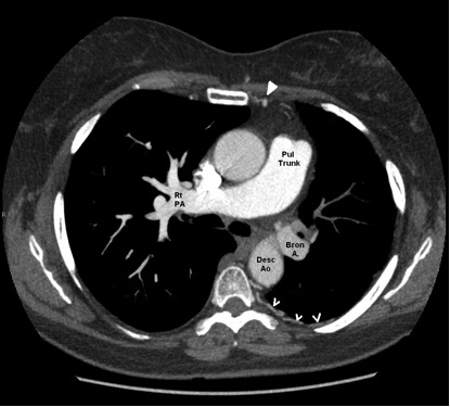
The CT image is an axial slice of the thorax from a CT pulmonary angiogram study as evidenced by the dense enhancement of the pulmonary artery.

The right pulmonary artery courses in direct continuity from the pulmonary trunk, however the left pulmonary artery is not seen connecting with the pulmonary trunk.  In addition to this, the vessel in the region of the left pulmonary artery is poorly enhanced compared with the right pulmonary artery, and matches the enhancement seen in the descending aorta.  The combination of these features is in keeping with isolated unilateral absence of the pulmonary artery (IUAPA)2 with compensatory dilatation of the bronchial artery arising from the descending aorta.  As well as the dilated bronchial artery, the left internal mammary artery and intercostal arteries are also dilated, acting as further sources of collateral supply to the left lung.  The left lung is small compared with the right due to a degree of pulmonary atresia (Figure 2).

Figure 2: Annotated figure demonstrating the variant anatomy of this case of Isolated unilateral absence of a pulmonary artery

IOTM May 2018 fig2

Pul Trunk = Pulmonary trunk, Rt PAS = right pulmonary artery, Desc Ao = descending aorta, Bron A = Bronchial artery.  Filled in arrowhead points to a dilated Left internal mammary artery (cf the other side), Open arrows point to dilated intercostal arteries.


Q3: What complications is this patient at risk of?

  1. Left-sided pulmonary hypertension
  2. Right sided bronchiectasis
  3. Left sided haemoptysis
  4. Pulmonary embolism

Recurrent infections, dyspnoea and decreased exercise tolerance are the most common presentations of this condition.2  In addition to this there are several important sequalae.  Due to the increased blood through the single remaining pulmonary artery these patients are at high risk for developing pulmonary artery hypertension, this is especially pronounced during pregnancy, and indeed this can be the trigger that leads to the condition being discovered.4  Reduced blood supply to the side with the absent pulmonary artery predisposes it to recurrent infections with reports of bronchiectasis secondary to this.The dilated bronchial artery, internal mammary arteries and intercostal arteries are all at increased risk of bleeding due to the increased pressures running through them and their abnormal tortuous nature.  Patients with IUAPA are also at increased risk for high altitude pulmonary oedema and should be warned as such.  UAPA can occur with other congenital anomalies (in which case it is no longer “isolated”), in particular ASDs, VSDs and Tetralogy of Fallot.  Echocardiography is a useful adjunct in investigation as it can assess for these at the same time as measuring for pulmonary artery pressures to diagnose pulmonary hypertension.

References:

  1. Tsoumakidou MChrysofakis GTsiligianni IMaltezakis GSiafakas NMTzanakis N.  A prospective analysis of 184 hemoptysis cases: diagnostic impact of chest X-ray, computed tomography, bronchoscopyRespiration. 2006;73(6):808-14. Epub 2006 Jan 27.
  2. Ten Harkel ADBlom NAOttenkamp J. Isolated unilateral absence of a pulmonary artery: a case report and review of the literature. Chest. 2002 Oct;122(4):1471-7.
  3. Yiu MWC, Le DV, Leung Y, Ooi CGC.  Radiological features of isolated unilateral absence of the pulmonary artery.  J HK Coll Radiol 2001;4:277-280.
  4. Stiller RJ, Soberman S, Turetsky A, et al.  Agenesis of the pulmonary artery,: an unusual cause of dyspnea in pregnancy.  Am J Obstet Gynecol 1988; 158:172-173.

Image of the Month – March 2018 – Progressive dyspnoea in a patient with a bioprosthetic AVR

Progressive dyspnoea in a patient with a bioprosthetic AVR

Jack PM Andrews, Marc R Dweck, Alastair J Moss.

Centre for Cardiovascular Science, University of Edinburgh, Chancellor’s Building, Royal Infirmary of Edinburgh, 51 Little France Crescent, Edinburgh EH16 4TJ, UK.

Corresponding Author: Dr. Jack PM Andrews. E-mail: Jack.Andrews@ed.ac.uk

Introduction

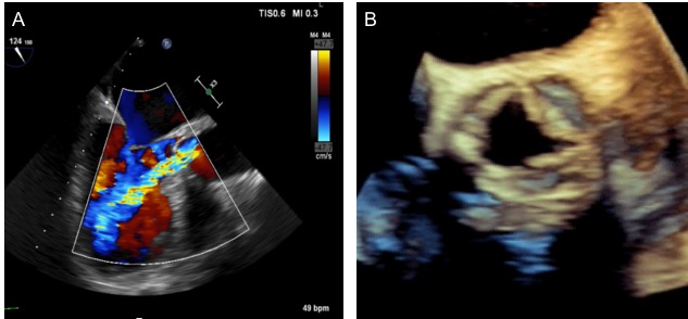
An 85-year old woman presented to the cardiology service with exertional dyspnoea whilst walking on the flat. She underwent a bioprosthetic surgical aortic valve replacement (stentless 25 mm Kohler Elan, Vascuetec) 12 years ago for calcific aortic stenosis. She was otherwise systemically well and had not undergone any recent dental procedures. On examination, she had a collapsing pulse (BP 120/30 mmHg) and grade 3 early diastolic murmur in the aortic area. Electrocardiogram (ECG) demonstrated sinus rhythm with voltage criteria for left ventricular hypertrophy and routine bloods returned a normocytic anaemia with a mildly elevated serum bilirubin (27 umol/L). A transoesophageal echocardiogram (TOE) was performed. (Figure 1)

Figure 1. Transoeophageal echocardiogram mid-oesophageal AV long axis view (A). 3D echocardiogram short axis view (B).


Figure 2. Cardiac magnetic resonance demonstrated transprosthesis laminar regurgitation jet (A). 3D  cardiac computed tomography coronal reconstruction of the aortic bioprosthesis confirmed the left cusp prolapse [inset] that is independent from a septated LVOT aneurysm (B). Pre TAVR deployment aortogram showing substantial volume of contrast within the left ventricle confirming significant aortic incompetence (C). Post TAVR deployment aortogram; note the lack of contrast within the left ventricle indicating the return of valvular competence and successful TAVR deployment (D).

Clinical Question

What is the aetiology of the bioprosthetic aortic valve dysfunction?

  1. Infective endocarditis
  2. Late suture dehiscence
  3. Leaflet prolapse
  4. Thrombus formation
  5. LVOT aneurysm formation

Answer – C

The correct answer is leaflet prolapse. TOE demonstrated a severe central regurgitant jet at the level of the prosthesis, confirmed by the large width of the colour flow jet (>65% of LVOT diameter) and colour M-mode holodiastolic flow reversal across the prosthesis (Figure 1A & B). Pathological regurgitation of prosthetic heart valves can either be central or paravalvular. Paravalvular regurgitation is often related to position of the sewing ring and anchoring tissue in the outflow tract with latent failure of the suture line due to age-related deterioration or LVOT aneurysm formation. Central regurgitation is a common feature of structural valve dysfunction. Distortions in leaflet anatomy due to perforations or thickening of the cusps ultimately result in leaflets prolapsing below the annular plane.

Whilst echocardiography remains the initial first line investigation, adjuvant imaging using cardiac magnetic resonance is increasingly utilised to confirm the severity and location of regurgitant jets (Figure 2A). [1] Bioprosthetic valve thrombosis and infective endocarditis are important differential diagnoses to exclude using modalities with superior spatial resolution such as cardiac computed tomography (Figure 2B). [2]

Figure 2C and 2D demonstrate aortograms both pre and post deployment of transcathether aortic valve replacement (TAVR) illustrating the immediate cessation of valvular incompetence.  The advent of valve-in-valve interventions offers novel strategies for high-risk patients with failing stentless bioprostheses. [3] Confirming the specific aetiology of bioprosthesis dysfunction with multi-modality imaging is a priority to select the most appropriate therapeutic strategy. This patient underwent transcatheter aortic valve implantation (26mm CoreValve Evolut R, Medtronic) with an immediate improvement in symptoms.

REFERENCES

[1]       Salaun E, Jacquier A, Theron A, Giorgi R, Lambert M, Jaussaud N, Hubert S, Collart F, Bonnet JL, Habib G, Cuisset T, Grisoli D. Value of CMR in quantification of paravalvular aortic regurgitation after TAVI. Eur Heart J – Cardiovascular Imaging. 2016;17:41-50.

[2]       Moss AJ, Dweck MR, Dreisbach JG, Williams MC, Mak SM, Cartlidge T, Nicol ED, Morgan-Hughes GJ. Complementary role of cardiac CT in the assessment of aortic valve replacement dysfunction. Open Heart. 2016;3:e000494. Doi: 10.1136/openhrt-2016-000494.

[3]       Duncan A, Davies S, Di Mario C, Moat N. Valve-in-valve transcatheter aortic valve implantation for failing surgical aortic stentless bioprosthetic valves: A single-center experience. J Thorac Cardiovasc Surg. 2015;150:91-98.

Image of the Month – February 2018 – An unusual cause of chest pain

An unusual cause of chest pain

Dylan Yong, Nadir Khan, Michelle Williams

Royal Infirmary of Edinburgh, University of Edinburgh

Case history

A 78-year-old female presented with chest pain of increasing frequency.  She had undergone investigation of chest pain with invasive coronary angiography and balloon angioplasty to her left coronary artery 2 weeks prior.

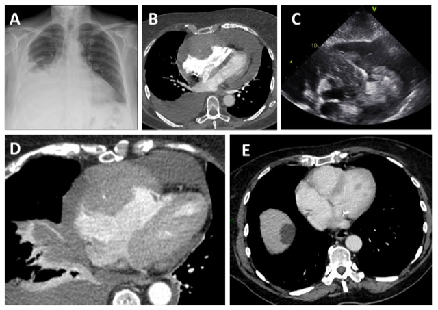
Admission chest x-ray showed a right sided pleural effusion and an enlarged cardiac contour (Figure 1A). Her troponin and D-dimer were raised. Electrocardiogram (ECG) was unremarkable.  She underwent a CT pulmonary angiogram (CTPA) which demonstrated a large pericardial effusion with maximum depth of 3.7cm (Figure 1B). Within this, there was an area of homogenous high attenuation in the epicardial space, following the course of right coronary artery. This was causing mass effect on the adjacent right atrium and ventricle. There was evidence of cardiac tamponade with straightening of the interventricular septum. Echocardiogram revealed a mass in the epicardial space suspicious for malignancy (Figure 1C).

A therapeutic pericardial drain was performed and haemoserous fluid obtained was sent for cytology. A total of 1.5 litres of fluid were drained from the right pleural space. Repeat CT post pericardial drainage demonstrated the mass more clearly (Figure 1D). Cytology from the pericardial effusion confirmed a diagnosis of primary cardiac diffuse large B cell lymphoma, stage IV. CT staging demonstrated no other sites of disease. Following discussion in a multi-disciplinary meeting, she received R-CHOP chemotherapy. She had an excellent response to the treatment, with a repeat 3-month interval CT scan demonstrating no measurable disease. (Figure 1E)

Discussion

Cardiac malignancy is rare, and primary cardiac lymphoma (PCL) accounts for only 1% of primary cardiac tumours.1 Amongst the small number of cases of PCL both B-cell and T-cell lymphomas have been reported.1 The majority of the PCL arise from the right side of the heart, particularly the right atrium.2 The reason for this distribution is unknown2. Cardiac involvement may also occur in up to 20% of patients who have diffuse nodal lymphoma.Lymphoma is an important differential diagnosis for cases when more than one cardiac chamber is involved.

Diagnosis of primary cardiac tumours is typically late and they are associated with a poor prognosis.2Symptoms are related to the site of involvement of the heart. Ikeda et al. reviewed the presentations of 40 patients with PCL and found that patients generally complain of non-specific symptoms such as dyspnoea and oedema, or present with arrhythmias and pericardial effusion.3

Figure Legend

Figure 1A. Chest X-ray with an enlarged cardiac contour and a right pleural effusion.

Figure 1B. CTPA showing a right pleural effusion and pericardial effusion. Homogenous material surrounding the right coronary artery causing mass effect on the right atrium and right ventricle.

Figure 1C. Transthoracic echocardiography showing a mass suspicious for malignancy in the epicardial space.

Figure 1D. CT of the chest after pericardial drainage showing the mass in the epicardial space.

Figure 1E. CT of the chest after treatment showing no evidence of residual disease.

Multiple-choice questions

1. What percentage of patients with diffuse nodal lymphoma have cardiac involvement?

  1. 5%
  2. 10%
  3. 20%
  4. 30%
  5. 40%

2. What is the name given to the appearance of a pericardial effusion on chest x-ray?

  1. Sillhouette sign
  2. Chang sign
  3. Water-bottle sign
  4. Double bubble sign
  5. Snowman sign

3. Which chemotherapy drug is correctly associated with the type of cardiovascular toxicity?

  1. Anthracycline – heart failure
  2. Cyclophosphamide – ischaemia
  3. Fluorouracil – hypertension
  4. Cisplastin – ischaemia, thromboembolism
  5. Vincristine – heart failure

Answers: 1/C, 2/C, 3/A

References

  1. Patel J, Melly L, Sheppard MN. Primar cardiac lymphoma: B- and T-cell cases at a specialist UK centre. Ann Oncol 2010; 21(5):1041-5
  2. Ceresoli GL, Ferreri AJ, Bucci E, Ripa C, Ponzoni M, Villa E. Primary cardiac lymphoma in immunocompetent patients: diagnostic and therapeutic management. Cancer 1997;80: 1497-1506.
  3. Ikeda H, Nakamura S, Nishimaki H, Masuda K, Takeo T, Kasai K, et al. Primary lymphoma of the heart: case report and literature review. Pathol Int 2004;54(3):187-95

Image of the Month – January 2018 – Pulmonary Vein Stenosis

Single narrow left sided stenotic pulmonary vein leading to pleural effusion and pulmonary congestion

Vicky Tilliridou, Michelle C Williams.

Royal Infirmary of Edinburgh, Edinburgh, United Kingdom.


Case Report

A 42-year-old gentleman with paroxysmal atrial fibrillation underwent multiple electrophysiological interventions for symptoms refractory to medication. These included several radiofrequency ablations and one cryoablation for pulmonary vein isolation. Three weeks post-cryoablation, he reported right-sided non-exertional chest pains lasting for up to 5 minutes per episode with associated chronic cough and exertional breathlessness.

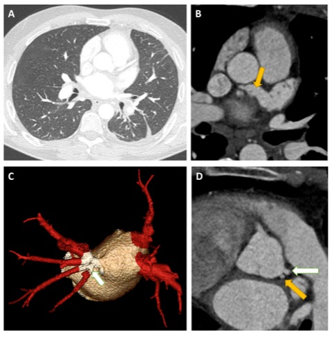
Computed tomography coronary angiography (CTCA) was performed to evaluate the pulmonary venous anatomy. Four pulmonary veins were visualised draining together into the left atrium with stenosis of the left lower pulmonary vein. Wide field-of-view images demonstrated features of pulmonary venous congestion and a small left pleural effusion. Additionally a large, patent “chicken wing” atrial appendage was present. Incidentally, the left coronary artery had separate origins from the left coronary cups for the left anterior descending artery and left circumflex artery.

Figure: A. Wide field-of-view image showing evidence of cardiac failure (increased fluid in the fissures) and a left pleural effusion. B. Axial image showing a very large left atrial appendage with a “chicken wing” configuration (yellow arrow). C. 3D reconstruction showing two pulmonary veins on the right and one pulmonary vein on the left with a stenotic narrowing at the ostium. D. Separate origins of the left anterior descending and left circumflex artery from the left coronary cusp.

Discussion

Radiofrequency catheter ablation was introduced in the 1980s as a novel treatment method for atrial fibrillation. It was initially performed within the pulmonary vein itself but this commonly led to pulmonary vein stenosis. The technique evolved to performing circumferential ablation in the cardiac antrum around the ostia of the pulmonary veins has become the standard approach with a reduced long-term risk of pulmonary vein stenosis (1). In the most widely used definition, from the HRS/EHRA/ECAS Expert Consensus Statement, mild stenosis is present if the reduction compared to pre-ablation diameter is < 50%, moderate if 50-70%, and severe if ≥ 70% (2). Recent data shows that acquired pulmonary veins stenosis secondary to catheter ablation for atrial fibrillation occurs in a mean of 2% and a median of 3.1% of cases (3). However, the incidence following ablation for atrial fibrillation may be underestimated as patients may be asymptomatic or may remain unrecognised due to non-specific symptoms.  Indeed, although symptoms may include chest pain, breathlessness, cough, or haemoptysis, patients often remain asymptomatic if the collateral circulation compensates for the reduced flow through the narrowed pulmonary vein(s).

Plain radiography and CT imaging may show pulmonary venous congestion or pleural effusions, however these are non-specific signs of left-sided heart failure. Diagnosis of pulmonary vein stenosis can be achieved with CT angiography, magnetic resonance imaging, transoesophageal echocardiography or catheter venography (4-6).

Multiple choice questions

1. In what proportion of the population is the typical pattern of four pulmonary veins and four well-differentiated ostia seen? (7)

A) <50%

B) 60-70%

C) 70-80%

D) 80-90%

E) >90%


2. Which is the most common focus of atrial fibrillation? (7)

A) Left superior pulmonary vein

B) Left inferior pulmonary vein

C) Right superior pulmonary vein

D) Right inferior pulmonary vein


3. Which imaging modality can identify and quantify pulmonary vein stenosis?

A) Plain radiography

B) CT angiography

C) MR perfusion

D) Single photon emission tomography


Answers

1 = B

2 = A

3 = B


References

1.         Edriss H, Denega T, Test V, Nugent K. Pulmonary vein stenosis complicating radiofrequency catheter ablation for atrial fibrillation: A literature review. Respir Med. 2016;117:215-22.

2.         Calkins H, Kuck KH, Cappato R, Brugada J, Camm AJ, Chen SA, et al. 2012 HRS/EHRA/ECAS Expert Consensus Statement on Catheter and Surgical Ablation of Atrial Fibrillation: recommendations for patient selection, procedural techniques, patient management and follow-up, definitions, endpoints, and research trial design. Europace. 2012;14(4):528-606.

3.         Pazos-Lopez P, Garcia-Rodriguez C, Guitian-Gonzalez A, Paredes-Galan E, Alvarez-Moure MA, Rodriguez-Alvarez M, et al. Pulmonary vein stenosis: Etiology, diagnosis and management. World J Cardiol. 2016;8(1):81-8.

4.         Schneider C, Ernst S, Bahlmann E, Malisius R, Krumsdorf U, Boczor S, et al. Transesophageal echocardiography: a screening method for pulmonary vein stenosis after catheter ablation of atrial fibrillation. Eur J Echocardiogr. 2006;7(6):447-56.

5.         Scharf C, Sneider M, Case I, Chugh A, Lai SW, Pelosi F, Jr., et al. Anatomy of the pulmonary veins in patients with atrial fibrillation and effects of segmental ostial ablation analyzed by computed tomography. J Cardiovasc Electrophysiol. 2003;14(2):150-5.

6.         Yang M, Akbari H, Reddy GP, Higgins CB. Identification of pulmonary vein stenosis after radiofrequency ablation for atrial fibrillation using MRI. J Comput Assist Tomogr. 2001;25(1):34-5.

7.         Porres DV, Morenza OP, Pallisa E, Roque A, Andreu J, Martinez M. Learning from the pulmonary veins. Radiographics. 2013;33(4):999-1022.

Image of the Month – December 2017 – Multimodality imaging in a case of left atrial myxoma

Multimodality imaging in a case of left atrial myxoma

Williams, MG1; Lewis, K2; MacIver, DH3; Sheffield, E4, Gosling, O3

  1. Department of Cardiology, Bristol Heart Institute, Bristol, UK
  2. Department of Radiology, Musgrove Park Hospital, Taunton, UK
  3. Department of Cardiology, Musgrove Park Hospital, Taunton, UK
  4. Department of Pathology, North Bristol NHS Trust, Bristol, UK

An asymptomatic 52-year-old gentleman was referred to our district general hospital for routine review due to a family history of aortopathy. He had no past medical history and was normotensive. An initial transthoracic echocardiogram (TTE) revealed normal left ventricular function and normal indexed aortic root dimensions.

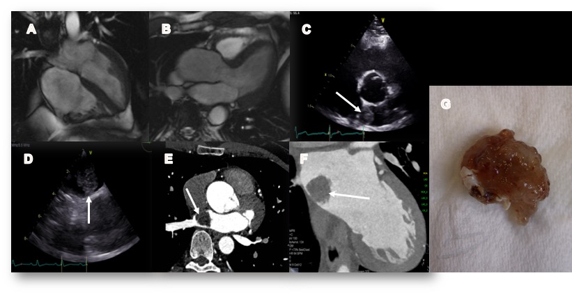
He returned for a follow up scan 5 years later and he was found to have asymptomatic moderate LV systolic dysfunction on TTE. Cardiovascular magnetic resonance imaging (CMR) was arranged. This confirmed moderate left ventricular impairment (ejection fraction 43%), no regional wall motion abnormality, no late gadolinium enhancement, a bicuspid aortic valve and a mildly dilated aortic root (Figure 1A-B). There was no evidence of an intra or extra-cardiac mass. The patient was commenced on a beta-blocker and angiotensin receptor blocker.

Two years later (Figure 1C) a repeat TTE now demonstrated a large left atrial mass. He was admitted and a transoesophageal echocardiogram (Figure 1D) and a cardiac CT (Figure 1E-F) were performed which confirmed the presence of a 22 x 15 mm pedunculated left atrial mass attached to the interatrial septum. There was no evidence of metastases. The CT also confirmed normal coronary arteries.  He was discussed with our local cardiothoracic centre and was transferred urgently for surgical resection (Figure 1G). He has made an excellent recovery and subsequent histological analysis confirmed this was a myxoma.


Figure 1:  4 (A) and 3 chamber (B) cardiac MRI images showing dilated aortic root and no evidence of atrial myxoma in 2015. C Subsequent transthoracic echocardiogram (2017) in the parasternal short axis showing echogenic lesion (white arrow) in the left atrium. D Transoesophageal echocardiogram (TOE) demonstrating the pedunculated lesion attached to the interatrial septum. Contrast-enhanced coronary CT angiogram showing the mass in the axial (E) and oblique (F) views. (G) Photograph of excised lesion prior to histological analysis


Multiple choice questions:

1.  What is the median age of presentation of a myxoma?

A) 20-30 years

B)  30-40 years

C)  40-50 years
D) 50-60 years

E)  60-70 years

Answer C)

Cardiac myxomas are seen most frequently in adult females. At presentation, the median age is 49 years. However, cases have been reported in patients under 20 years and over 90 years of age1.  

2. Which statement regarding myxoma is correct?

A) Myxoma is the most common type of tumour found in the heart.

B) Treatment is urgent  due to high risk of fatal embolism and haemodynamic collapse

C)  Local recurrence is common and affects >20% of cases

D) Roughly half of myxomas are solitary

E) The right atrium is the most common location

Answer B)

Metastatic tumours are 20-40 times more common than myxomas, which are the most common primary cardiac tumour2. Surgical excision should be considered in all patients as a matter of urgency. Death from embolic complications between diagnosis and treatment has been estimated at between 8-10%3.  Local  recurrence  is rare affecting 3-4% of patients4. 90% of myxomas are solitary and the left atrium is the most common location representing 86% of myxomas1.  

 3. Which of the following statements regarding the appearance of a myxoma on CT is false?

A) They demonstrate high attenuation  on non-contrast CT

B) 10% are calcified

C) Pedunculated appearance

D) Heterogeneous contrast enhancement

E) May be seen to prolapse through the mitral valve

Answer A)

Myxomas usually exhibit low attenuation on non-contrast CT. 10% may be calcified, more commonly when they are located in the right atrium. They are often pedunculated and attached to the interatrial septum via a stalk. They enhance heterogeneously due to the individual tumours’ chronicity and degree of necrosis and haemorrhage. Large tumours may prolapse through the mitral valve and can cause LV inflow obstruction2.


References:

1Mir IA et al. Atrial myxoma ‘a review’ International journal of community medicine and public health 2016 Jan; 3(1):23-29

2Kassop D et al. Cardiac Masses on Cardiac CT: A Review. Current Cardiovascular Imaging Reports. 2014;7(8):9281. doi:10.1007/s12410-014-9281-1.

3Dato GMA et al.  Long term follow up of cardiac myxoma, (7-31years). Journal of Cardiovascular Surgery. 1993;34(2):114-43.

4Castells E et al. Cardiac myxoma: Surgical treatment, long term results and recurrence. J Cardiovasc Surg. 1993;34(1):49-53.

2Kassop D et al. Cardiac Masses on Cardiac CT: A Review. Current Cardiovascular Imaging Reports. 2014;7(8):9281. doi:10.1007/s12410-014-9281-1.

3Dato GMA et al.  Long term follow up of cardiac myxoma, (7-31years). Journal of Cardiovascular Surgery. 1993;34(2):114-43.

4Castells E et al. Cardiac myxoma: Surgical treatment, long term results and recurrence. J Cardiovasc Surg. 1993;34(1):49-53.

Image of the Month – November 2017 – An 8 month old child presenting with ischaemic cardiomyopathy

Case Study: an 8 month old child presenting with ischaemic cardiomyopathy

Matt Muller, Philip Gonsalves Radiology SpRs – Newcastle Hospitals

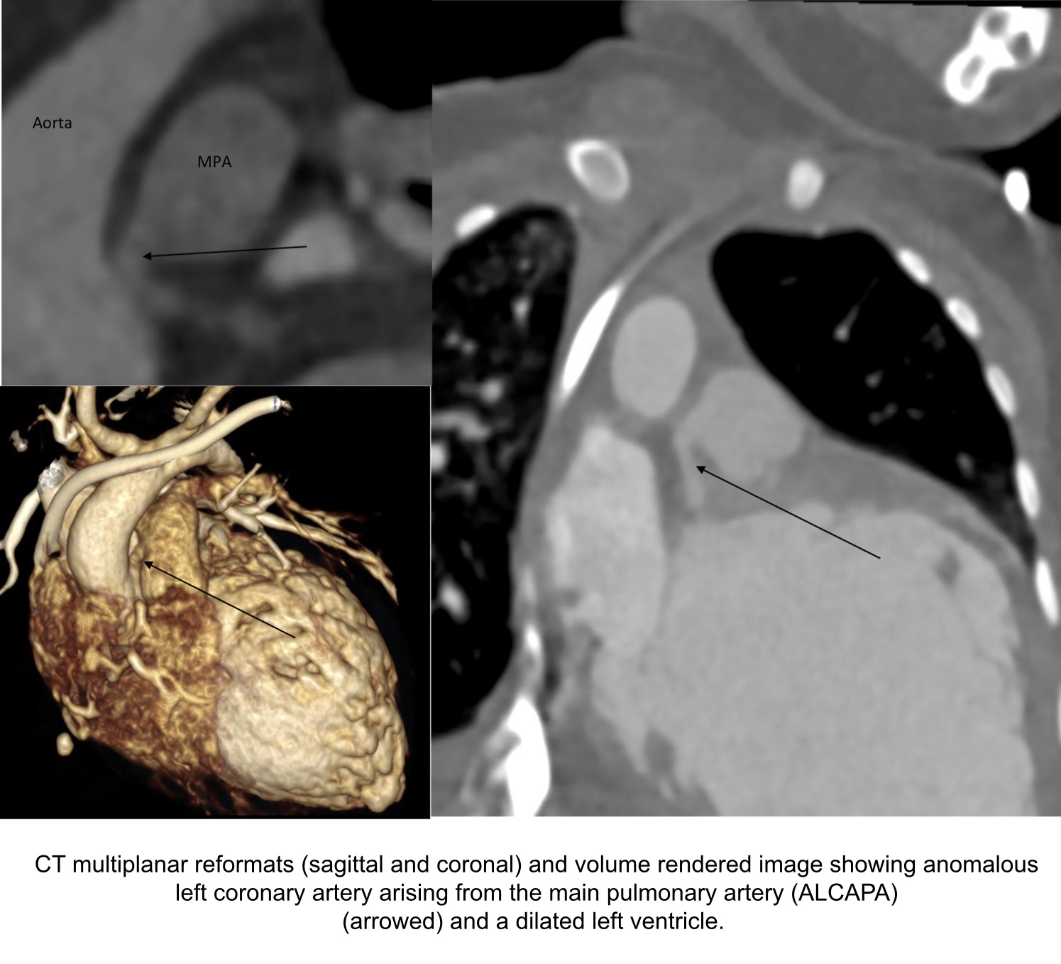
An 8-month old male born at term with known VACTERL association presented to his local hospital and a diagnosis of ischaemic cardiomyopathy and heart failure was made. Comorbidities included an imperforate anus, tracheoesophageal fistula, horseshoe kidney and nephrocalcinosis. The infant was transferred to the Freeman Hospital in Newcastle upon Tyne for further management of his severe cardiomyopathy of unknown etiology. The ECG appeared ischaemic. An echocardiogram revealed a dilated left ventricle with global left ventricular impairment and an ejection fraction of 27%. It was not possible to view the origin of the LAD at echocardiography. A CT coronary angiogram was performed which confirmed an anomalous left coronary artery arising from the pulmonary artery (ALCAPA). In utero, the pulmonary arterial and systemic arterial pressures are equal. Therefore, there is antegrade flow in both the normal RCA and the ALCAPA. After birth, the pulmonary arterial pressure and oxygen saturation drops, which results in the left ventricle being perfused by relatively desaturated blood under low pressure. Therefore, myocardial ischemia ensues [1]. The patient underwent cardiac surgery where the ALCAPA was re-implanted into the aorta. The patient was then transferred back to their base hospital for further management.


Questions

  1. The acronym VACTERL stands for vertebral defects, anorectal anomalies, cardiac defects, tracheoesophageal fistula, renal anomalies and limb anomalies. How many characteristic features need to be present to make the diagnosis?
  2. a)3
  3. b)4
  4. c)6

Answer – typically 3 features need to be present [2].

  1. What ECG findings are associated with ALCAPA?
  2. Widespread concave ST elevation
  3. LBBB and reduced voltage QRS complexes
  4. Abnormal Q waves in leads I, avL, V5, and V6, as well as by transient ST changes in these leads

 Answer – The ECG findings associated with ALCAPA usually shows typical signs of an anterolateral myocardial infarction, manifested by abnormal Q waves in leads I, avL, V5, and V6, as well as by transient ST changes in these leads. 20-45% of patients do not show abnormal Q waves so the diagnosis should be strongly suspected if there is abnormal R wave progression in the chest leads [3].

References

[1] https://emedicine.medscape.com/article/893290-overview#a5

[2] https://ghr.nlm.nih.gov/condition/vacterl-association

[3] Electrocardiogram of anomalous left coronary artery from the pulmonary artery in infants. Hoffman JI, Paediatric Cardiology 2013 Mar;34(3):489-91

Our Sponsors