Image of the Month – July 2016 – An unusual cause of a left to right shunt

Dr Jonathan Rodrigues, Bristol Heart Institute

Case:

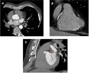
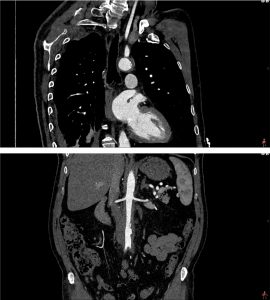
An elderly gentleman presented with chest pain, shortness of breath and haemoptysis. His past medical history was significant for previous type A aortic dissection and ascending aortic repair many years ago. On examination, he was hypoxic and cyanosed. An urgent, non-ECG gated CT aorta was performed. This revealed a large pseudoaneurysm arising from the superior aspect of the ascending aortic repair (Figure 1A and 1B). The pseudoaneurysm was in direct continuity with the right main pulmonary artery (Figure 1A-C). There was evidence of significant left to right shunt from ascending aorta, via the pseudoaneurysm, into the right-sided pulmonary arterial system, with differential opacification of the main pulmonary arteries, with avid right-sided enhancement on the systemic arterial phase study (Figure 1A and 1C). The discrepancy in size of segmental and subsegmental pulmonary arteries, larger in the right lower lobe compared to the left, was suggestive of acute right-sided pulmonary arterial volume loading (Figure 2A). The airspace opacification in the middle and right lower lobes (Figure 2B) was consistent with alveolar oedema and possibly haemorrhage from rapid significantly increased hydrostatic pressure and possibility capillary haemorrhage due to the large volume acute shunt. Unfortunately, the patient deteriorated very quickly and did not survive for definitive surgical correction to be attempted.

Discussion:

Acquired aorto-pulmonary artery fistulation is a rare but life-threatening condition. It may arise as a complication of aortic aneurysms[1] and both acute and chronic aortic dissections[2]. However, progression of a post-aortic surgery pseudoaneurym to an acquired aorto-pulmonary fistula is exceedingly rare. Potential causes for such a rare complication are postulated to include longstanding right pulmonary arterial compression by the pseudoaneurysm, atherosclerosis, connective tissue disorders, necrosis, trauma, inflammatory aortitis and infection [3]. Aorto-pulmonary fistulae can be asymptomatic [4] or more commonly present with congestive heart failure, chest pain and haemoptysis [5]. Definitive treatment of aorto-pulmonary fistulae is usually surgery. However, treatment with Amplatzer Septal Occluder has previous been described [6].

Figure 1. Non-ECG gated arterial phase CT of the thorax.

A) Axial image showing a large ascending aortic pseudoaneurysm (§) exerting mass effect on the ascending aortic graft repair (*), from previous type A aortic dissection. There is a direct connection between the ascending aortic pseudoaneurysm and the right main pulmonary artery (solid white arrow). There is differential opacification of the right and left main pulmonary arteries with more avid enhancement in the right main pulmonary artery (dotted white arrow) compared to the left main pulmonary artery (dashed white arrow) on this arterial phase study.

B) Sagittal reformatted image showing the ascending aortic graft repair (*) with large pseudoaneurysm arising from the proximal aspect of the graft repair (§). The connection between the pseudoaneurysm and the right main pulmonary artery is demonstrated (solid white arrow).

C) Coronal reformatted image showing an abnormal serpiginous connection with the right main pulmonary artery (solid white arrow) and differential opacification of the main pulmonary arteries on this systemic arterial phase study, with more avid opacification of the right main pulmonary artery (dotted white arrow) compared to the left main pulmonary artery (dashed white arrow).
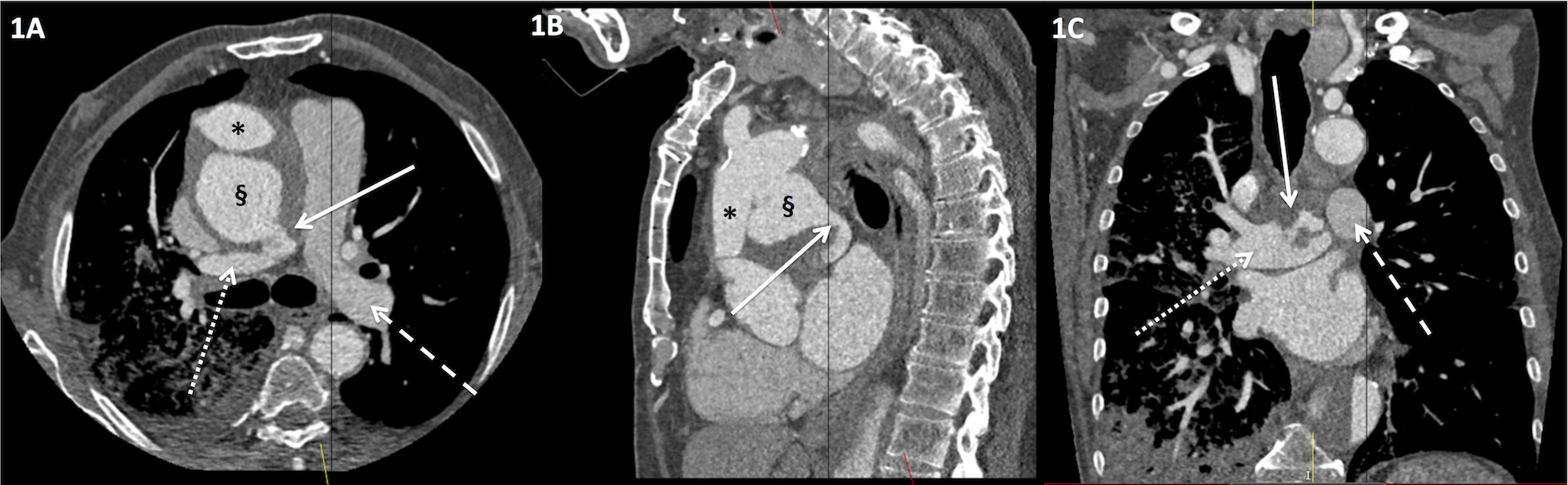
Figure 2. Non-ECG gated arterial phase CT of the thorax

A) Axial image display on mediastinal window settings showing asymmetrical enlargement of the right basal segmental and subsegmental pulmonary arteries (dotted white arrow) compared to the left basal pulmonary arteries (dashed white arrow). There is evidence of previous dissection in the descending thoracic aorta with opacified oval-shaped true lumen (black *) and thrombosed false lumen (white *).

B) Axial image displayed on lung window settings showing focal consolidation in the right lower lobe (*) and the lateral segment of the middle lobe. In this clinical context, this was consistent with alveolar haemorrhage.

References

[1] Boyd LJ. A study of four thousand reported cases of aneurysm of the thoracic aorta. Am J Med Sci. 1924; 168: 654-663.

[2] Piciche M, De Paulis R, Chiariello L. A review of aortopulmonary fistulas in aortic dissection. Ann Thorac Surg. 1999; 68: 1833-6.

[3] Mukadam M, Barraclough J, Riley P, et al. Acquired aorto-pulmonary artery fistula following proximal aortic surgery. Interact Cardiovasc Thorac Surg. 2005; 4: 388-90.

[3] Maeder MT, Wobler T, Kunzli A et al. Aortopulmonary fistula occurring 4 years after replacement of the ascending aorta. Ann Thorac Surg. 2006; 81: e18-e20.

[4] Ferrari G, Anastasio G, Bianchi M et al. Aortopulmonary fistula after a modified bentall procedure. J Heart Valve Dis. 2012; 21: 505-508.

[5] Wakefield BJ, Winter D, Alfirevic A. Staged repair of an aortopulmonary fistula from a large ascending aortic pseudoaneurysm: the role of transesophageal echocardiography. J Cardiothorac Vasc Anesth. 2015. doi: 10.1053/j.jvca.2015.11.022. [Epub ahead of print].

[6] Coserria F, Mendez A, Moruno A, et al. Percutaneous closure of iatrogenic aortopulmonary fistula using the Amplatzer Septal Occluder. Rev Esp Cardiol. 2014: 67: 228-229.
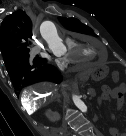
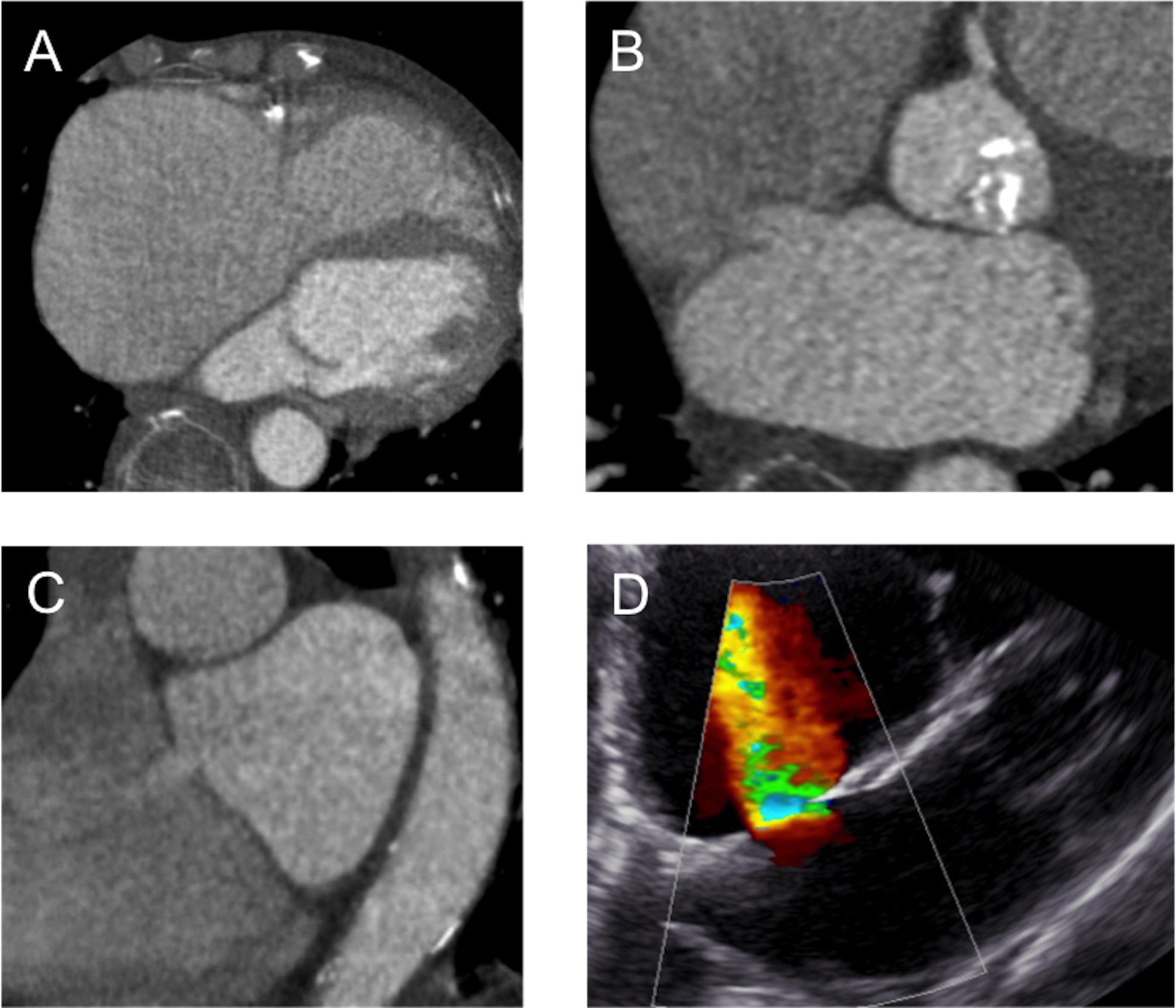
Image of the Month – June 2016 – Acute aortic syndrome in extremis

Dr. John Dreisbach, Glasgow Royal Infirmary

Introduction

The term acute aortic syndrome covers a spectrum of life-threatening emergency conditions of the thoracic aorta, including dissection, intramural haematoma and penetrating atherosclerotic ulcer. The potential acute complications are severe and diverse. The clinical manifestations are often non-specific and overlap significantly with other more common conditions requiring essentially the opposite treatment, most frequently acute coronary syndrome and pulmonary embolism. The best chances of survival depend on a rapid and accurate diagnosis (or indeed exclusion), usually by CT angiography and ideally cardiac-gated to minimise motion artifact.

Questions

Review the single image below (a coronal-oblique reconstruction of a CT angiogram) and address:

  1. The type of acute aortic syndrome present
  2. The Stanford classification
  3. The major complication that has occurred
  4. The signs that this major complication is life-threatening


Answers

    1. Acute intramural haematoma of the ascending aorta

There is an area of eccentric high-attenuation lining the wall of the ascending aorta, without discernible enhancement, consistent with an acute intramural haematoma.

    2. Stanford type A

The Stanford classification of aortic dissections aids decision-making for management and also broadly applies to intramural haematomas. Type A dissections involve the ascending aorta and should be managed surgically whereas type B dissections classically commence distal to the left subclavian artery and should be managed medically.

The Stanford classification system is not without many important exceptions and caveats, but is generally useful and routinely employed in both clinical practice and research.

    3. Large haemopericardium

There is a large pericardial collection of high attenuation consistent with acute haemopericardium, in this case due to rupture of the intramural haematoma of the ascending aorta.

Haemopericardium is the commonest complication of both type A aortic dissections and intramural haematomas and the most frequent cause of death in the acute setting, the mechanism of which is described below.

    4. Cardiac tamponade (also known as pericardial tamponade), in this case evidenced by a large volume of relatively undiluted intravenous contrast material in a dependent position within the hepatic veins of the right liver lobe

The accumulation of fluid (in this case haemorrhage) into the pericardial sac, which is lined by a stiff fibrous capsule, transmits pressure onto the underlying heart. This can cause compression and reduced filling of the cardiac chambers, resulting in reduced cardiac output and obstructive shock.

The slow accumulation of fluid may allow time for the pericardium to stretch and accommodate relatively large-volume effusions before causing hemodynamic compromise. However, only a small volume (100-200 ml) of rapidly accumulating pericardial fluid may be required to produce obstructive shock. Unfortunately this patient suffered the worst of both – the rapid accumulation of a large pericardial collection.

In this patient, intravenous contrast was injected into an upper limb vein and undiluted contrast is seen in transit in the SVC. A large volume of undiluted intravenous contrast has travelled down the SVC, bypassed the right atrium, refluxed down into the IVC, and produced a striking appearance by accumulating in a dependent position within the hepatic veins and venous tributaries of the right liver lobe. The reflux of intravenous contrast into the IVC and dilated hepatic veins are key CT signs that the right heart is failing to fill with venous return and in this case represents severe cardiac tamponade at the time of the examination.

References

  1. Multidetector CT of aortic dissection: a pictorial review. McMahon MA, Squirrell CA. RadioGraphics 2010;30(2):445–460. http://pubs.rsna.org/doi/full/10.1148/rg.302095104
  1. Recommendations for accurate CT diagnosis of suspected acute aortic syndrome (AAS)—on behalf of the British Society of Cardiovascular Imaging (BSCI)/British Society of Cardiovascular CT (BSCCT). Varut Vardhanabhuti, Edward Nicol, Gareth Morgan-Hughes, Carl A Roobottom, Giles Roditi, Mark C K Hamilton, Russell K Bull, Franchesca Pugliese, Michelle C Williams, James Stirrup, Simon Padley, Andrew Taylor, L Ceri Davies, Roger Bury, and Stephen Harden. The British Journal of Radiology 2016 89:1061. http://www.birpublications.org/doi/abs/10.1259/bjr.20150705

Image of the Month – April 2016 – Mind the gap: atrial septal defects in the elderly

An 86 year-old male presented with progressive shortness of breath on exertion on a background of severe aortic stenosis. Cardiac computed tomgraphy demonstrated a hypertrophied left ventricle with a severely dilated right atrium (Figure A). Axial and sagittal multi-planar reconstructions revealed an ostium secundum atrial septal defect (ASD) with transit of contrast from the left to right atrium (Figure B and C). Transthoracic echocardiography confirmed a left-right shunt through the ostium secundum ASD (Figure D).

Ostium secundum ASDs are the most common type of ASD accounting for 80-90% of defects in the interatrial septum [1]. Shunt flow occurs predominantly during ventricular diastole with the direction of flow determined by difference in the compliance and capacity between the left and right ventricles [2]. Cardiac computed tomography acquisitions during late-diastole with opacification of the left heart can visualise ASDs without the requirement for the Valsalva manoeuvre.

In elderly patients with ostium secundum ASDs, larger left-right shunts may occur following changes in left ventricular compliance. In this case, the shunt acts as a ‘release valve’ for the left atrium to maintain lower pulmonary venous pressures. Closure of the defect may lead to an acute rise in left atrial pressure and precipitate pulmonary oedema.

References

[1] Johri AM, Rojas CA, El-Sherief A, et al. Imaging of the atrial septal defects: echocardiography and CT correlation. Heart. 2011;97:1441-1453.

[2] Sommer RJ, Hijazi ZM, Rhodes Jr. JF. Pathophysiology of Congenital Heart Disease in Adults. Part I: Shunt Lesions. Circulation. 2008;117:1090-1099.

Image of the Month – March 2016 – single coronary artery arising from the right coronary cusp

Submitted by Dr I. Nagra, Consultant Radiologist, Worcestershire Acute Hospital.

Coronary artery anomalies (CAAs) are present in 1-1.96% of the population (1,2).  The significance of single CAA’s differs depending on the course of the artery.  Those that take an inter-arterial route between the aorta and pulmonary artery are considered malignant, often presenting with syncope or sudden death (1,3). This is attributed to myocardial ischaemia from compression of the single coronary artery (SCA) between two high-flow structures in systole. Conversely, those that traverse benign, inter-ventricular paths do not predispose to myocardial ischaemia and hence are often asymptomatic (1,3).

The CT coronary angiography and volume rendered images shows a SCA arising from the right coronary cusp in a 68 year-old male who presented with atypical chest pain. The images show both the right coronary artery (RCA) and left main stem (LMS) arising from the right coronary cusp. A long benign, inter-ventricular course of the LMS is undertaken before bifurcation into the LAD and LCx, both of which contain calcified eccentric plaques. Mixed plaque disease causing moderate to severe stenosis of the RCA was noted. This CAA is rare, with a prevalence of 0.004 – 0.05% (2). According to the Lipton classification system, this represents an R-II S single coronary artery anomaly (4).

Given the benign course of the SCA, the atypical chest pain is likely to be secondary to the significant plaque disease rather than the CAA. Some authors suggest that atherosclerosis is accelerated in those with a SAA (5). Knowledge of anomalous coronary artery anatomy is of considerable benefit especially if invasive treatment for coronary artery disease is required.


References

  1. Laspas F, Roussakis A, Mourmouris C, Kritikos N, Efthimiadou R, Andreou J. Coronary artery anomalies in adults: imaging at dual source CT coronary angiography. J Med Imaging Radiat Oncol. 2013 Apr;57(2):184-90.
  1. Erol C, Seker M. Coronary artery anomalies: the prevalence of origination, course, and termination anomalies of coronary arteries detected by 64-detector computed tomography coronary angiography. J Comput Assist Tomogr. 2011 Sep-Oct;35(5):618-24.
  1. Angelini P, Flamm SD. Newer concepts for imaging anomalous aortic origin of the coronary arteries in adults. Catheter Cardiovasc Interv. 2007 Jun 1;69(7):942-54.
  1. Lipton MJ, Barry WH, Obrez I, Silverman JF, Wexler L. Isolated single coronary artery: diagnosis, angiographic classification, and clinical significance. Radiology. 1979;130:39-47.
  1. Rigatelli G, Gemelli M, Zamboni A, Docali G, Rossi P, Rossi D, et al. Are coronary artery anomal

Image of the Month – February 2016 – A 6 year old boy with swallowing difficulties

Submitted by Dr Michelle Mak

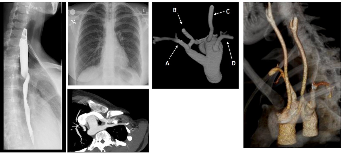
A 6 years old boy presented with swallowing difficulties. His chest radiograph is as shown. A barium swallow showed a posterior indentation, and a CT was requested.

Questions:

  1. Given the MIP MPRs, name the arrowed vessels A-D.
  2. What is this vascular anomaly?


Answers:

  1. A: Left subclavian artery (LSCA).

B: Left common carotid artery (LCCA)

C: Right common carotid artery (RCCA)

D: Right subclavian artery (RSCA).

2.  Right aortic arch (RAA) with aberrant LSCA.

There are multiple arch anomalies, and they can be detected prenatally on foetal ultrasound, and postnatally with both ECHO and cross sectional imaging. A vascular ring typically encircles the trachea and oesophagus completely. RAA is present in 0.05% of the population. Based on the embryonic double aortic arch model, a RAA with aberrant LSCA is due to an interruption between the LCCA and LSCA. The first branch of the arch is the LCCA, followed by the RCCA and RSCA. Lastly, the LSCA is aberrant. If the LSCA origin is dilated, it is known as Kommerell’s diverticulum. The ring is commonly completed by a left ligamentum arteriosum, which could be treated by surgical ligation.

The second commonest pattern of RAA exhibits a mirror image branching pattern, with the left brachiocephalic artery arising first, followed by the right common carotid and right subclavian artery. It is commonly associated with congenital heart diseases and is asymptomatic.

References

  1. Donnelly L, Fleck R, Pacharn P et al. Aberrant Subclavian Arteries: Cross-Sectional Imaging Findings in Infants and Children Referred for Evaluation of Extrinsic Airway Compression. AJR:178, May 2002
  2. Türkvatan A, Büyükbayraktar G, Ölçer T et al. Congenital Anomalies of the Aortic Arch: Evaluation with the Use of Multidetector Computed Tomography. Korean J Radiol. 2009 Mar-Apr; 10(2): 176–184.

Image of the Month – January 2016 – Acute onset chest pain and ST elevation

Submitted by Dr Yan Ning Neo, Dr Jonathan Weir-McCall
Ninewells Hospital and Dundee Medical School, UK

This 61 year old male presented with acute onset chest pain, and now onset ST elevation on his ECG on a background of prior left circumflex stenting several years previously for stable angina.  He was immediately transferred to the cath lab where engagement of the left coronary was technically challenging.  After stenting of the LMS, angiography continued to show marked irregularity of the left circumflex artery and complete occlusion of the LAD, and a markedly abnormal appearance of the aortic sinus.  In addition the right coronary artery could not be cannulated.  The suspicion of an aortic dissection was raised and the patient was transferred straight from the cath lab for an emergency ungated CT.

The short axis view of the left ventricle (Figure A) demonstrates, even on this ungated exam, a transmural perfusion defect involving the anterior and septal walls consistent with LAD occlusion. Oblique axial (Figure B), and sagittal images (Figure C) of the aorta root show a highly unusual circumferential aortic dissection which involves both coronary ostia with the dissection flap intussuscepting into the ascending aorta.  Note is also made of the intra-aortic balloon pump on the oblique sagittal view (Figure C) where the CO2 filled tube is inflating and deflating during image acquisition.

Diagnosis: Circumferential ascending aorta dissection with intimal intussusception with extension of the dissection into the left coronary artery causing a left anterior descending artery (LAD) infarct.

Circumferential intimal dissection as seen in this case is a rare but hazardous complication of aortic dissection, and is termed “intimo-intimal intussusception”.[1] Complications of this can arise due to the dissection flap extending into vessels such as the coronary ostia, or alternatively, unique to this kind of dissection, the cylindrical-shaped dissection flap can intussuscept proximally into the left ventricle outflow tract during diastole causing coronary ostia occlusion and potentially severe aortic valve insufficiency.[2] Complete or persistent coronary malperfusion can then lead to myocardial infarction as seen in the current case.[3]

References:

  1. Hufnagel CA, Conrad PW. Intimo-intimal intussusception in dissecting aneurysms. Am J Surg. 1962:103:727-31
  1. Whitley W, Tanaka KA, Chen EP et al. Acute aortic dissection with intimal layer prolapse into the left ventricle. Anesth Analg. 2007;104(4):774-6
  1. Lentini S, Sossio Perrotta. Aortic dissection with concomitant acute myocardial infarction: From diagnosis to management. J Emerg Trauma Shock. 2011;4(2):273-8.

Image of the Month – December 2015 – The Value of Iterative Model Reconstruction in cardiac and extra-cardiac findings

Submitted by Dr Michelle Crawford Jefferson

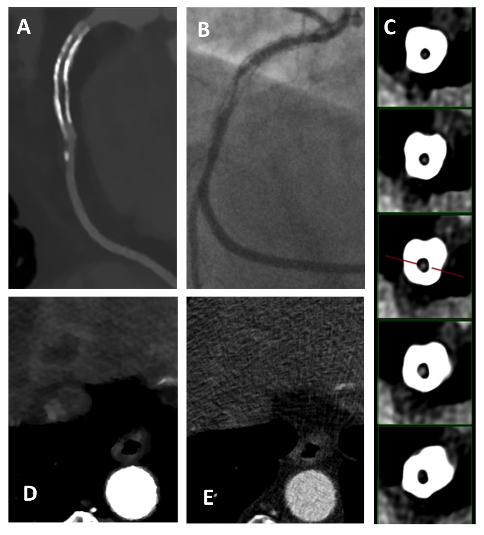
A 65 year old gentleman with previous RCA stent insertion was admitted with chest pain and anaemia and was referred for cardiac CT (Philips iCT 256 slice). The iMR (Iterative Model Reconstruction) reconstruction demonstrated in-stent restenosis in the proximal RCA (Image A, C) which was confirmed on catheter angiography (Image B). However it also showed a liver lesion (Image E) which was visible due to the improved contrast resolution with iMR, but not visible on the corresponding idose reconstruction (hybrid iterative reconstruction, Image E). A subsequent CT abdomen and CT colonography demonstrated a carcinoma within the descending colon and liver metastasis, which was the underlying cause of the anaemia.

Image of the Month – November 2015 – CT myocardial perfusion imaging to guide management

Submitted by Dr Michelle C Williams, Radiology ST3
Royal Infirmary of Edinburgh

CT myocardial perfusion imaging can provide information in addition to CT coronary angiography that can be used to guide patient management.1-3 Contrast enhanced electrocardiogram-gated CT images are acquired at rest and during pharmacological stress, such as with adenosine. Both “snap-shot” and “dynamic” CT protocols have been developed. This case shows an example where CT myocardial perfusion imaging was used to guide revascularization treatment.

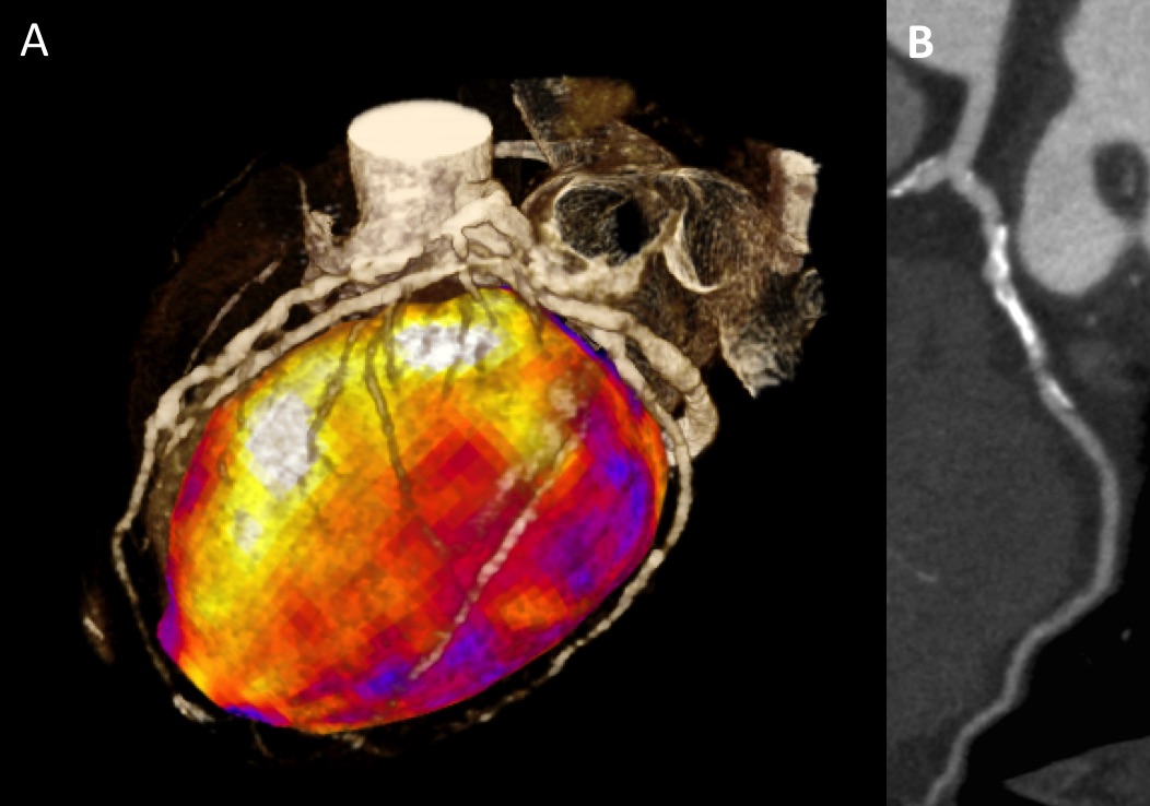
This male with known coronary artery disease presented with worsening symptoms of angina. CT coronary angiography identified heavily calcified atherosclerotic plaque in all three coronary arteries, any of which may have been the cause of his symptoms.  Figure A shows a three-dimensional reconstruction of the stress CT images with both the coronary arteries and myocardium. Multiple lesions in the coronary arteries can be identified. Figure B shows a curved planar reformation of the resting left circumflex artery, showing both calcified and non-calcified lesions. The patient underwent adenosine stress “snap-shot” CT myocardial perfusion imaging. In Figure A the myocardium is color-coded based on the attenuation density during stress imaging with white/yellow/orange/red showing normal perfusion and purple showing an area of reduced perfusion. This identified that the primary source of ischaemia was the left circumflex artery with a perfusion defect during stress imaging which resolved at rest. This meant that targeted revascularisation of the left circumflex artery could be performed.

REFERENCES
1.     Magalhães TA, Kishi S, George RT, et al. Combined coronary angiography and myocardial perfusion by computed tomography in the identification of flow-limiting stensois – The CORE320 study: An integrated analysis of CT coronary angiography and myocardial perfusion. J Cardiovasc Comput Tomogr. 2015.
2.     Rochitte CE, George RT, Chen MY, et al. Computed tomography angiography and perfusion to assess coronary artery stenosis causing perfusion defects by single photon emission computed tomography: the CORE320 study. European Heart Journal. 2014;35(17):1120-1130.
3.     Pelgrim GJ, Dorrius M, Xie X, et al. The dream of a one-stop-shop: Meta-analysis on myocardial perfusion CT. European Journal of Radiology. 2015.

Image of the month – October 2015 – A 46 year old lady with breathlessness

Submitted by Dr Alistair Moss
University of Edinburgh

A 46 year-old lady from South Africa was evaluated for breathlessness and heart failure. She had rheumatic fever aged two and aged nineteen she had severe calcific constrictive pericarditis and underwent 50% pericardiectomy. Transthoracic echocardiogram (TTE) revealed a calcified band with a calcific spur protruding through the anterolateral left ventricular (LV) wall, with tenting of the anterior mitral valve leaflet (Figure 1A Parasternal short axis and B modified apical 4 chamber TTE). CT coronary angiogram (CTCA) demonstrated a ring of dense calcification with LV impingement in two focal regions and systolic anterior motion of the anterior mitral valve leaflet. (Figure 1C Short axis CT and E 3D reconstruction of the calcification) The distal RCA was buried in the calcified pericardial band at the origin of the posterior descending artery (PDA) (Figure 1D, F & G).

QUESTION

What is the most likely aetiology of the calcified pericardial band?

  1. Previous cardiac surgery
  2. Radiotherapy
  3. Viral pericarditis
  4. Tuberculous pericarditis

ANSWER

The most likely cause of calcified pericardium (concretio cordis) in this case is silent tuberculous pericarditis. Pericardial constriction occurs in 20-50%. Calcified constrictive pericarditis can occur following cardiac surgery, but does not explain the initial presentation.  Chevers reported the first post-mortem case of calcified pericardial constriction in an 18 year-old female in 1841 as ‘the cavity of the pericardium was entirely obliterated by a layer of firm and almost cartilaginous deposit’.1 While case reports have identified pericardial calcification invading the right ventricular myocardium2, it is rare to find infiltration of the LV myocardium and coronary arteries.3

REFERENCES
1      Chevers N. Observations on the Diseases of the Orifice and Valves of the Aorta. Guy’s Hosp. Rep. 1842;7:387-442
2      Ahlgren B, Reece B, Salcedo E, Seres T. Constrictive pericarditis with a calcific mass invading into the right ventricular myocardium. Echocardiography 2013;30:E4-6
3      Gouley BA, Bellet S, McMillan TM. Tuberculosis of the Myocardium: Report of Six Cases with observations on involvement of the coronary arteries. Arch Int. Med. 1933;51:224

Image of the Month – September 2015 – 3D reconstruction of the coronary arteries

Submitted by – Dr Jason Sarfo-Annin CT2 Acute Care Common Stem,
Royal United Hospitals Bath NHS Foundation Trust

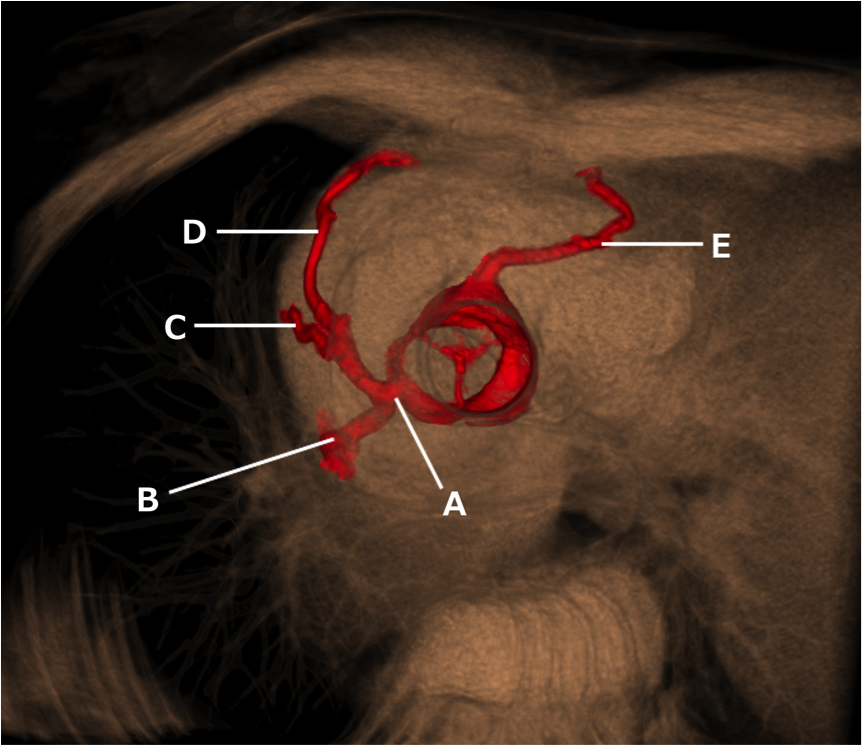
This three dimensional reconstruction of the chest shows the aortic valve and coronary arteries. Test yourself by naming the structures labelled A to E (Answers below the image).


Answers
A: Left main stem coronary artery
B: Left circumflex artery
C: First diagonal artery (D1)
D: Left anterior descending artery
E: Right circumflex artery

Our Sponsors