Submitted by Dr Michelle Crawford Jefferson
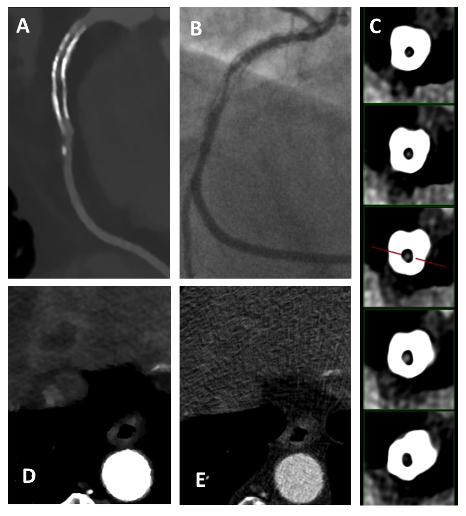
A 65 year old gentleman with previous RCA stent insertion was admitted with chest pain and anaemia and was referred for cardiac CT (Philips iCT 256 slice). The iMR (Iterative Model Reconstruction) reconstruction demonstrated in-stent restenosis in the proximal RCA (Image A, C) which was confirmed on catheter angiography (Image B). However it also showed a liver lesion (Image E) which was visible due to the improved contrast resolution with iMR, but not visible on the corresponding idose reconstruction (hybrid iterative reconstruction, Image E). A subsequent CT abdomen and CT colonography demonstrated a carcinoma within the descending colon and liver metastasis, which was the underlying cause of the anaemia.