Submitted by Dr Michelle C Williams, Radiology ST3
Royal Infirmary of Edinburgh
CT myocardial perfusion imaging can provide information in addition to CT coronary angiography that can be used to guide patient management.1-3 Contrast enhanced electrocardiogram-gated CT images are acquired at rest and during pharmacological stress, such as with adenosine. Both “snap-shot” and “dynamic” CT protocols have been developed. This case shows an example where CT myocardial perfusion imaging was used to guide revascularization treatment.
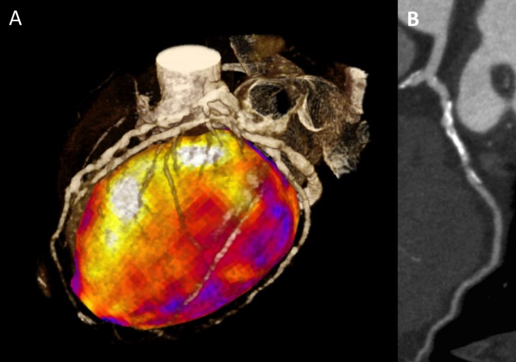
This male with known coronary artery disease presented with worsening symptoms of angina. CT coronary angiography identified heavily calcified atherosclerotic plaque in all three coronary arteries, any of which may have been the cause of his symptoms. Figure A shows a three-dimensional reconstruction of the stress CT images with both the coronary arteries and myocardium. Multiple lesions in the coronary arteries can be identified. Figure B shows a curved planar reformation of the resting left circumflex artery, showing both calcified and non-calcified lesions. The patient underwent adenosine stress “snap-shot” CT myocardial perfusion imaging. In Figure A the myocardium is color-coded based on the attenuation density during stress imaging with white/yellow/orange/red showing normal perfusion and purple showing an area of reduced perfusion. This identified that the primary source of ischaemia was the left circumflex artery with a perfusion defect during stress imaging which resolved at rest. This meant that targeted revascularisation of the left circumflex artery could be performed.
REFERENCES
1. Magalhães TA, Kishi S, George RT, et al. Combined coronary angiography and myocardial perfusion by computed tomography in the identification of flow-limiting stensois – The CORE320 study: An integrated analysis of CT coronary angiography and myocardial perfusion. J Cardiovasc Comput Tomogr. 2015.
2. Rochitte CE, George RT, Chen MY, et al. Computed tomography angiography and perfusion to assess coronary artery stenosis causing perfusion defects by single photon emission computed tomography: the CORE320 study. European Heart Journal. 2014;35(17):1120-1130.
3. Pelgrim GJ, Dorrius M, Xie X, et al. The dream of a one-stop-shop: Meta-analysis on myocardial perfusion CT. European Journal of Radiology. 2015.


