
Authors: Dr Rumneek Hampal, Dr David Gent
Case presentation
A gentleman in his 50s with cardiovascular risk factors including hypertension and raised BMI was transferred to his local PCI centre following an out of hospital VF arrest requiring 2 DCCV. ROSC was achieved following DCCV. On coronary angiography, there was a conus branch occlusion noted. A CMR performed during admission demonstrated impaired RV function, RV infarction with thrombus. The patient was treated with triple therapy (DOAC and dual antiplatelet therapy) with a plan for interval CMR in 3 months.
Key Imaging Findings
A cardiac MRI performed during admission demonstrated normal left ventricular size and wall thickness. Notably systolic function was preserved and there were no regional wall motion abnormalities. The right ventricle was severely dilated with moderate-severely impaired systolic function. There was akinesia of the mid-apical RV free wall and proximal RVOT. Basal segment function was preserved. RVEF was 37%.
Late gadolinium enhancement revealed extensive myocardial infarction of the mid to apical RV free wall extending to the proximal RVOT. A 2-4 mm layer of mural thrombus covered the infarcted segments on early and late enhancement images.
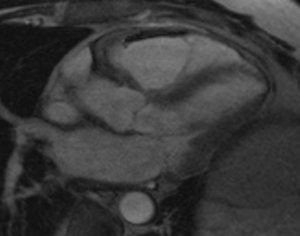
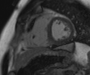
Early gadolinium enhancement imaging demonstrating; mural RV thrombus (arrow).
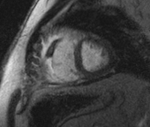
Delayed enhancement demonstrates transmural RV myocardial fibrosis
Discussion
This case describes out-of-hospital ventricular fibrillation arrest caused by isolated right ventricular infarction from occlusion of the conus branch of the right coronary artery. Conus occlusion is a recognised cause of malignant ventricular arrhythmia, but most reported cases are iatrogenic, occurring during catheter engagement or contrast injection during coronary angiography. Spontaneous occlusion presenting as cardiac arrest appears only in isolated case reports. The conus supplies the right ventricular outflow tract (RVOT), where ischaemia increases dispersion of repolarisation and creates a substrate for polymorphic VT/VF, a mechanism shared with Brugada syndrome (1). The diagnosis is easily missed. Right precordial ST elevation can mimic anterior left ventricular infarction, and the conus frequently arises from a separate ostium, so it may not opacify on selective right coronary injection.
Cardiac MRI was central to the diagnosis. Left ventricular size, thickness and function were normal, excluding a left ventricular infarct. The right ventricle was severely dilated (RVEF 37%), with infarction of the mid to apical free wall extending to the proximal RVOT. This extent, beyond the classical conus territory, may reflect a large conus branch or variant right ventricular supply. The overlying mural thrombus is notable. Post-infarction thrombus is described almost exclusively in the left ventricle, but the same mechanism is present in this case: akinetic myocardium, endocardial injury and a dilated, poorly contracting chamber.
Interval CMR will guide anticoagulation duration and assess right ventricular recovery.This case highlights that in VF arrest with anterior ST elevation and no left-sided culprit, the conus branch should be specifically sought.
Learning points
- Spontaneous conus branch occlusion is a rare but important cause of VF arrest and may be missed on angiography because the conus may have a separate ostium.
- Right precordial ST elevation with VF and no left-sided culprit should prompt evaluation for right ventricular or RVOT ischaemia.
- CMR can confirm isolated right ventricular infarction and identify complications such as mural thrombus.
- Right ventricular thrombus after right ventricular infarction is rare. Management is extrapolated from left ventricular thrombus practice and requires balancing thrombotic and bleeding risk on triple therapy.
References
- Ogano M, Iwasaki YK, Morita N, et al. Proarrhythmic ECG deterioration caused by myocardial ischemia of the conus branch artery in patients with a Brugada ECG pattern. Pacing Clin Electrophysiol. 2011;34(3):e26–e29. doi:10.1111/j.1540-8159.2010.02742.x.