Image of the Month April 2026: A case of Large Vessel Vasculitis with Coronary Artery Involvement.

Authors:

Dr Simon Woodbridge1, Dr Giulia Benedetti1


Affiliations:

1Guy’s & St Thomas’ NHS Foundation Trust


Case Presentation:

A 65 year old male presented following a 6 month history of progressively worsening exertional angina, on a background of unremarkable preliminary cardiac investigations, including normal electrocardiography. There was no history of exertional breathlessness or syncope. Other symptoms included occasional dizziness unrelated to posture or position, spontaneous shoulder cramping/stiffness, and occasional unilateral right sided headaches which would subside after approximately 1 hour.

His background medical history included previous tonsillar and prostate cancer, both in remission, and aside from being an ex-smoker, he otherwise demonstrated no risk factors for cardiovascular disease.


Imaging Findings:

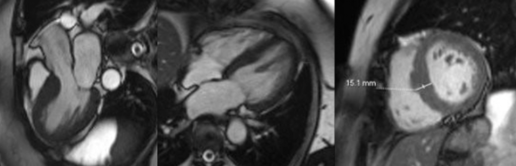
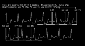
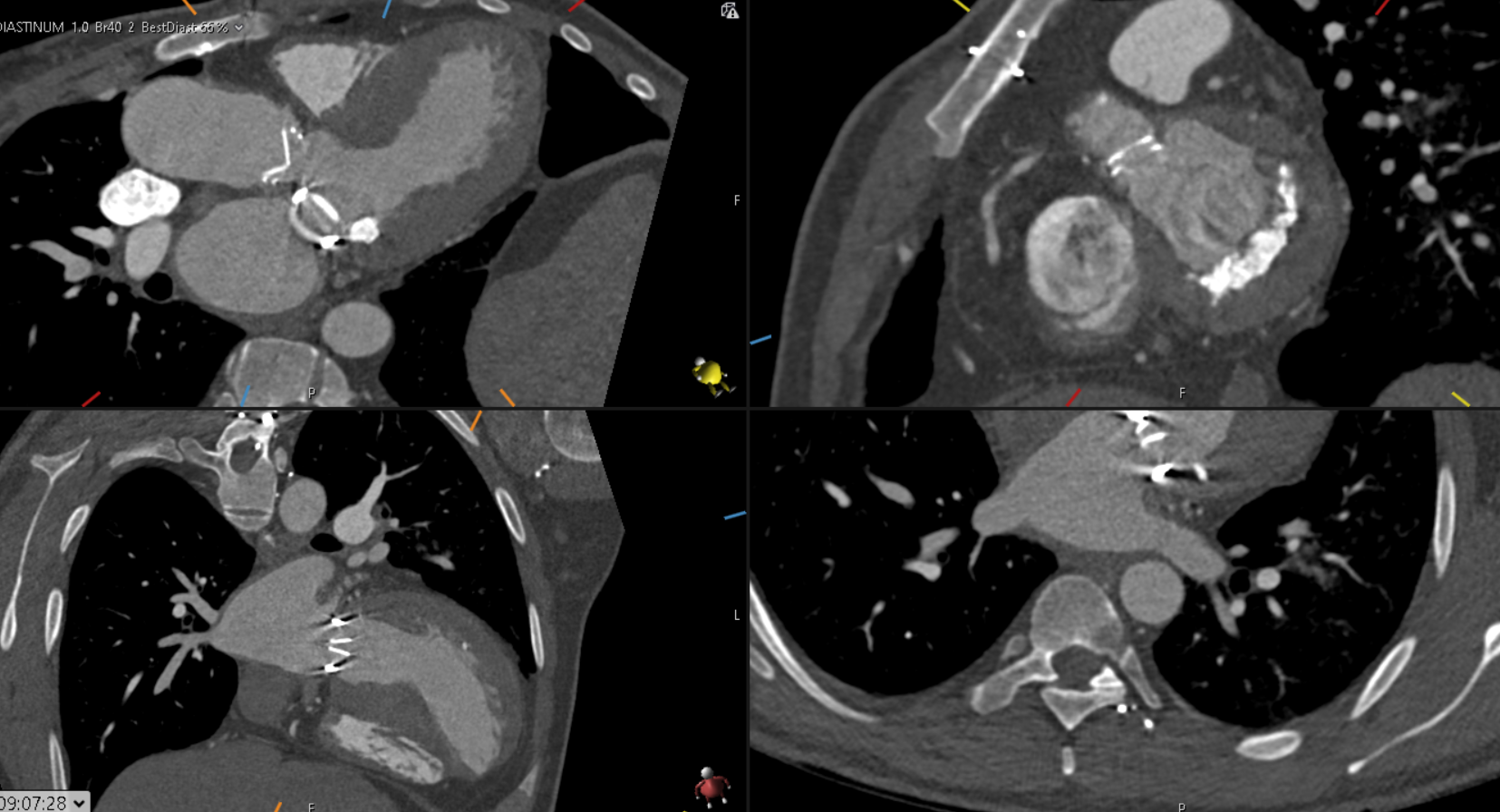
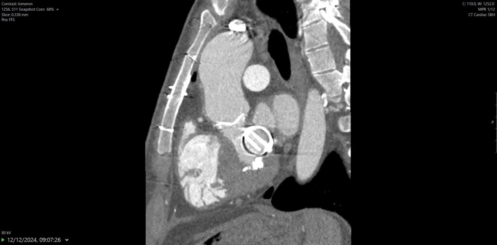
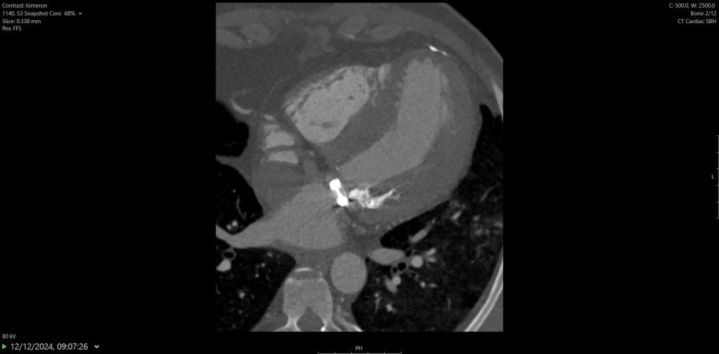
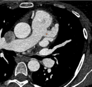
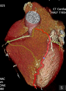
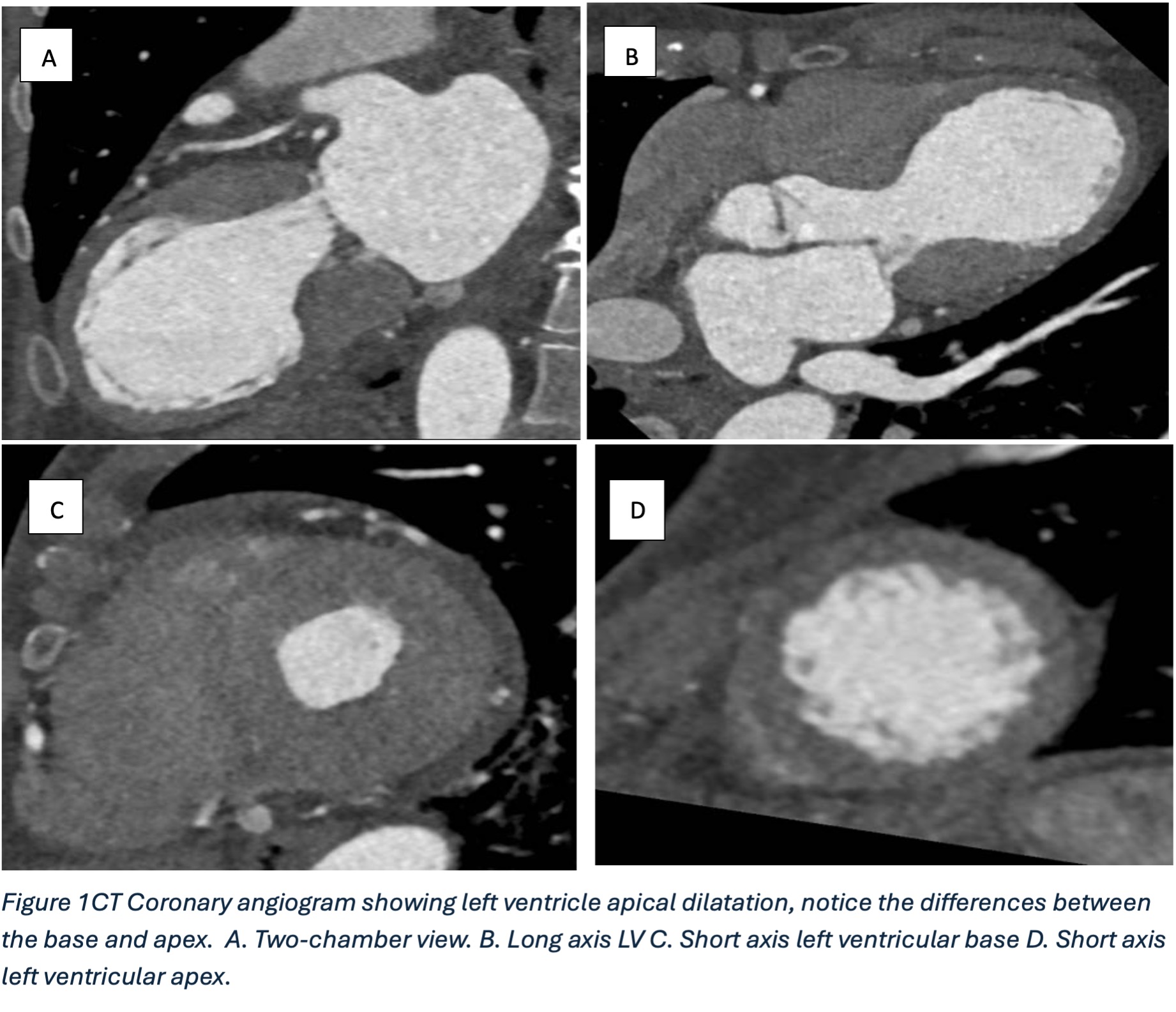
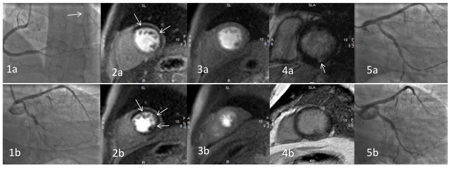
He was referred for CT coronary angiography to further investigate (Figure 1). This study revealed largely concentric perivascular soft tissue thickening affecting the left main stem, proximal left anterior descending (LAD) and left circumflex arteries, and proximal right coronary artery. At the affected areas, there was overall moderate (50-69%) stenosis. There was minimal calcific plaque within the proximal RCA.


Figure 1 | CT coronary angiogram demonstrating perivascular soft tissue thickening in the left main stem (A), proximal RCA (B) and proximal LAD (C)


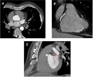
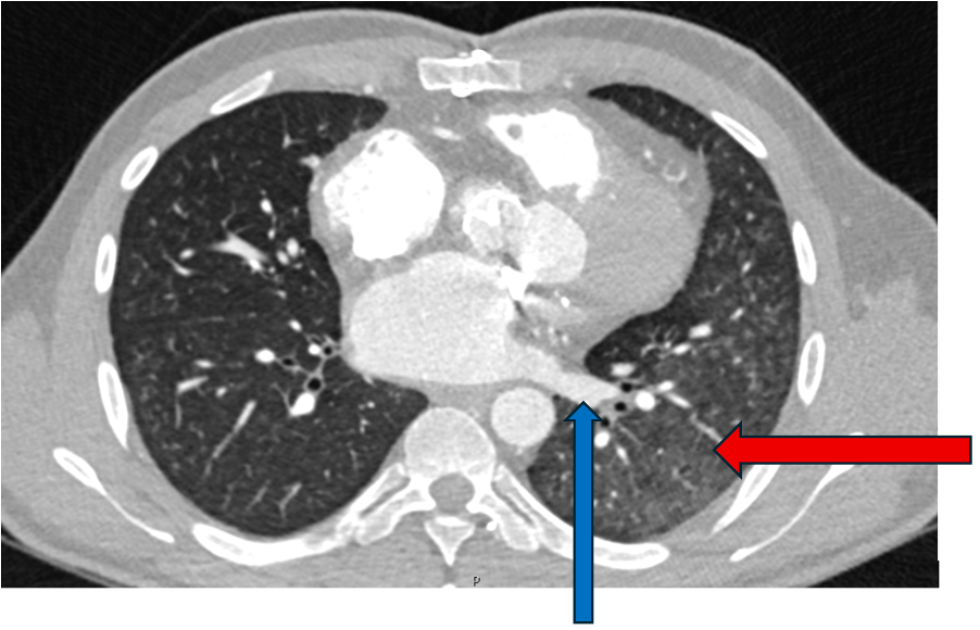
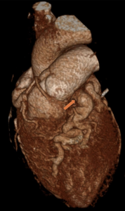
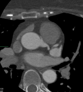
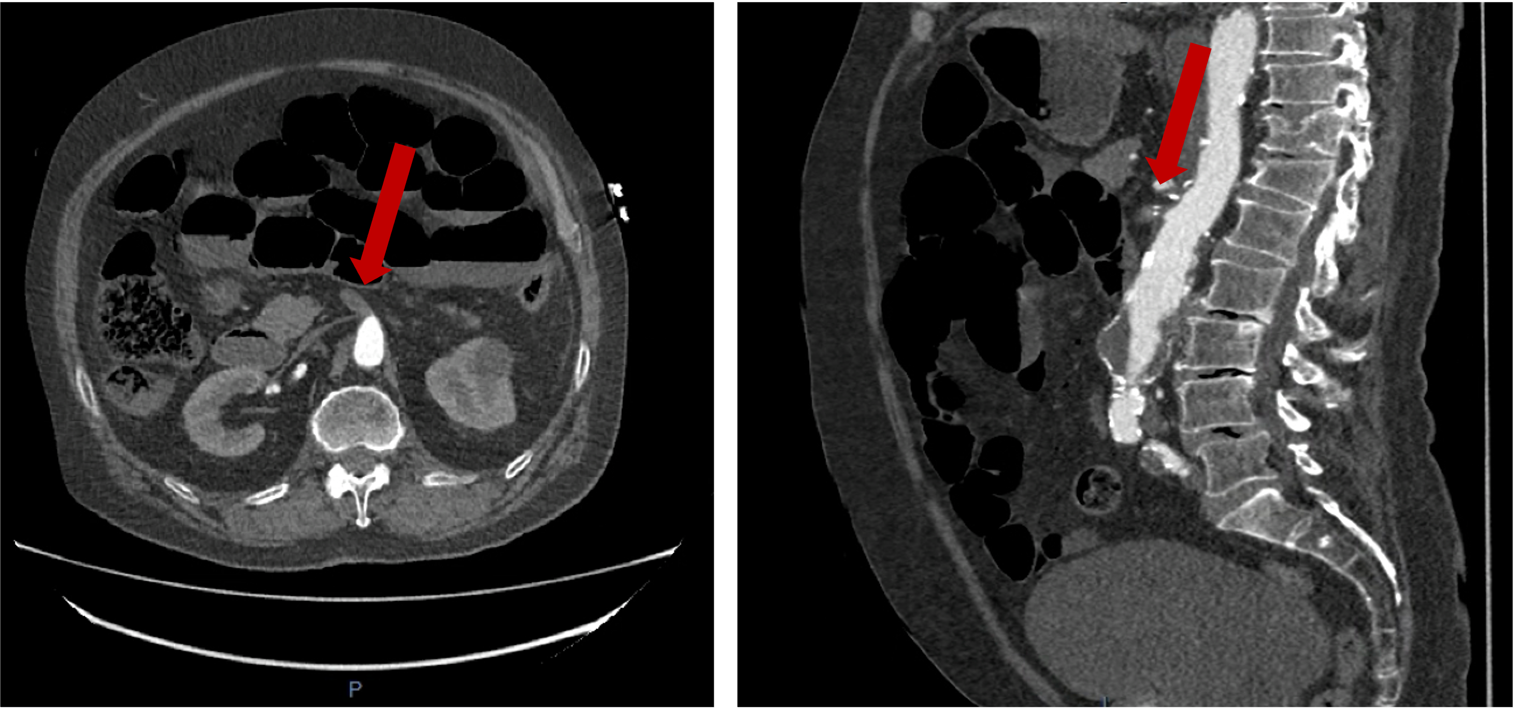
Due to the perivascular morphology of soft tissue thickening, rather than intraluminal plaque causing stenosis, in addition to the distribution of the abnormality predominantly within the proximal coronary vasculature, the possibility of vasculitis was raised, and further work-up was performed with a CT aortic angiogram (Figure 2). This study revealed circumferential aortic soft tissue thickening within the aortic arch, abdominal aorta, and proximal common iliac arteries. The remainder of the major aortic branch arteries, including the renal arteries, were spared. Suspicion of a vasculitic process was therefore increased.


Figure 2 | Coronal reformatting of CT aortic angiogram demonstrating peri-aortic soft tissue thickening, sparing the renal arteries.


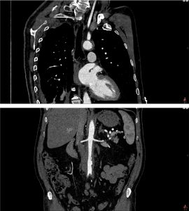
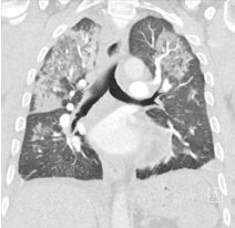
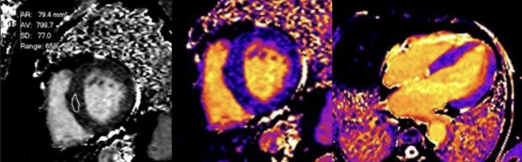
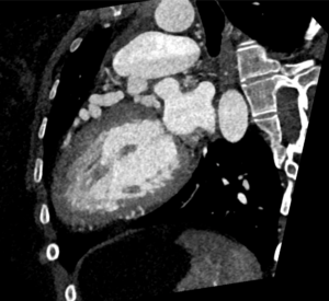
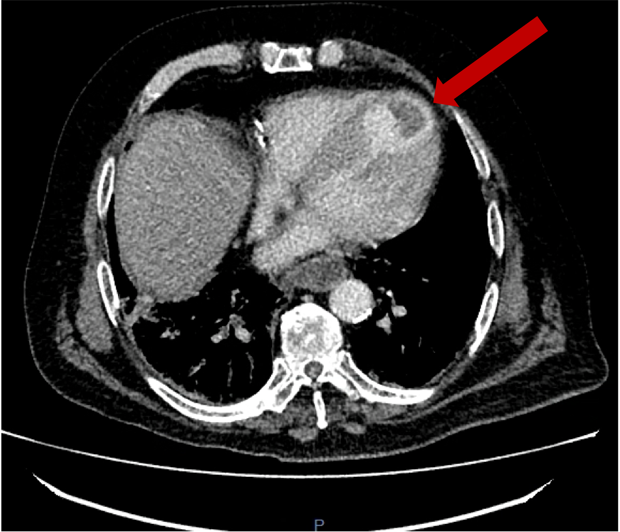
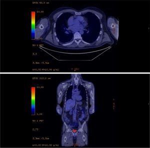
A subsequent FDG PET/CT study was performed (Figure 3). The PET/CT study demonstrated avid tracer uptake within the aorta and common iliac arteries at the sites of soft tissue thickening demonstrated on the prior CT aortic angiogram. In addition, a linear region of uptake correlating with the peri-coronary soft tissue along the LMS/LAD was identified, allowing for slight anatomical misalignment artefact and myocardial glucose utilisation. The distribution of inflammation, primarily centred around the coronary arteries, aorta and common iliac arteries, was radiologically deemed most in keeping with an IgG4 related vasculitis.


Figure 3 | FDG PET-CT demonstrating aortic/common iliac mural tracer uptake, as well as uptake within the region of the LMS/proximal LAD


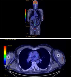
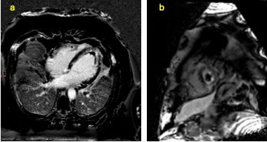
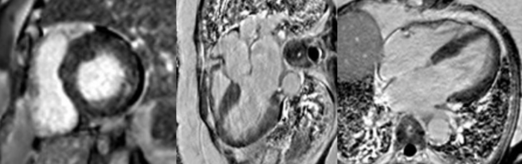
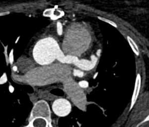
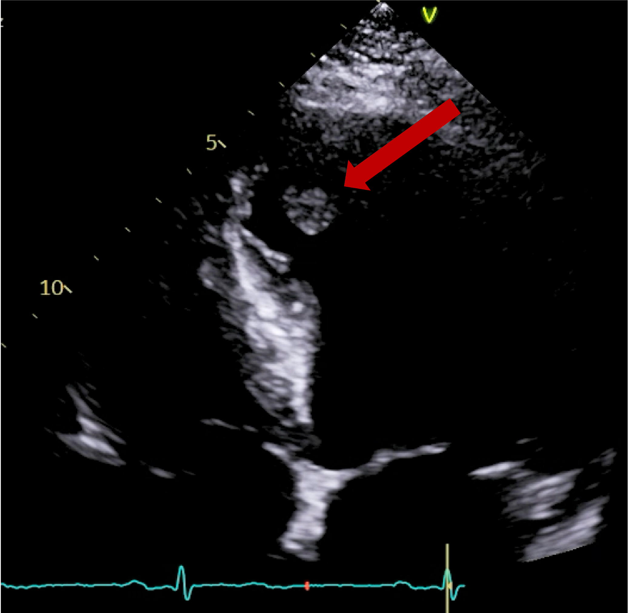
The patient was treated as a large vessel vasculitis, secondary to probable IgG4-related disease (serum IgG4 levels, reportedly not demonstrating high specificity in this condition, were normal) and commenced on high dose corticosteroid treatment. A repeat FDG PET/CT study was performed 1 month following treatment (Figure 4), which demonstrated a dramatic improvement, with minimal periaortic tracer uptake in the previously affected areas, in addition to complete cessation of uptake within the coronary arteries.


Figure 4 | Repeat FDG PET-CT demonstrating dramatic improvement/resolution at the previously demonstrated regions of tracer uptake in Figure 3.


Discussion:

IgG4 related disease (IgG4-RD) is a rare autoimmune systemic disease which is characterised by fibroinflammatory infiltration of organs by plasma cells expressing IgG4 antibodies. Elevated serum IgG levels are seen in 70-80% of patients, hence, normal levels do not exclude the disease.

There is a broad disease spectrum encompassing nearly all body systems and cardiovascular involvement can include aortitis, peri-aortitis, arteritis and coronary arteritis within its phenotype. In addition to coronary arterial and aortic inflammation, involvement of the common and external iliac arteries is the next most common affected vasculature, which was also exhibited in this patient.

CTCA, CTA and PET/CT imaging are key investigations which aid the diagnosis of IgG4 related cardiovascular disease. In CTA an additional delayed phase can assist in differentiating inflammatory perivascular thickening from mural thrombus formation if there is uncertainty in interpretation, showing possible contrast uptake within the soft tissue. In addition to diagnosis, PET/CT may also be a primary contributor in monitoring disease activity, as it is possible (as with this patient) that biochemical markers of inflammation do not correlate with disease extent.

Corticosteroids are the primary treatment of IgG4-RD, supplemented with methotrexate or mycophenolate mofetil/azathioprine as a second line option if there is limited response. A third line treatment option is B-cell depletion with Rituximab. This patient demonstrated a significant radiological and clinical response to corticosteroid treatment.


References:

Sudheer, N., Oliver, T.I. (2019). IgG4 Related Disease (IgG4-RD). [online] Nih.gov. Available at: https://www.ncbi.nlm.nih.gov/books/NBK499825/

Vasculitis UK. (n.d.). Immunoglobulin G4 Disease (IgG4-RD). [online] Available at: https://www.vasculitis.org.uk/about-vasculitis/immunoglobulin-g4

Oyama-Manabe, N., Satoshi, Y., Manabe, O., Kato, F., Hiromi Kanno-Okada and Kudo, K. (2018). IgG4-related Cardiovascular Disease from the Aorta to the Coronary Arteries: Multidetector CT and PET/CT. Radiographics, 38(7), pp.1934–1948. doi:https://doi.org/10.1148/rg.2018180049

Image of the Month March 2026: Clearer Vision, Safer Drainage!

Authors:

Shubham Garg1, Ruhani Bali1, Akash Batta1, Bishav Mohan1, Shitij Chaudhary1


Affiliation:

Department of Cardiology, Dayanand Medical College and Hospital, Ludhiana, India


Case Summary:

A 49-year-old male with no prior comorbidities presented to the emergency room (ER) with a 7-day history of fever, cough and shortness of breath. On admission, the patient had a blood pressure of 80/56 mmHg, a heart rate of 120 bpm, a respiratory rate of 36/minute, elevated jugular venous pressure, clear lung fields and muffled heart sounds on auscultation. An electrocardiogram was done, which showed sinus tachycardia with low-voltage QRS complexes. A screening bedside echocardiogram showed a large pericardial effusion (maximum thickness of 28 mm) with right ventricular diastolic collapse, suggestive of tamponade physiology. A bedside echocardiography-guided pericardiocentesis was attempted in the ER. Initially, 15 ml of haemorrhagic fluid was drained, after which no further fluid was drained. Cardiology consultation was obtained, and the patient was transferred to the cardiac catheterization laboratory.

Fluoroscopy was done, and the sheath was found to be in the splenic capsule (Figure 1). Under fluoroscopic guidance, a pigtail was inserted over a 0.035” J-tipped Terumo wire, after confirming the position of the wire in the pericardial space (Figure 2), and 250 ml of pericardial fluid was drained. Post-pericardiocentesis, the patient was vitally stable.


Figure 1 | 5F sheath inadvertently inserted into the splenic capsule during bedside pericardiocentesis.


Figure 2 | Fluoroscopic projection in AP view showing a pigtail catheter being introduced into the pericardial space over a 0.035-inch J-tipped wire. Note the characteristic path of the wire wrapping around the heart in the pericardial space.


Learning Points:

  1. Pericardiocentesis must always be done under fluoroscopic or expert echocardiographic guidance; blind procedures must be avoided.
  2. The position of the needle must always be confirmed once it enters the pericardial space.


Multiple Choice Questions:

  1. Which of the following clinical signs is not characteristic of cardiac tamponade?
    1. Jugular venous distension
    2. Muffled heart sounds
    3. Clear lung fields
    4. Bradycardia
    5. Pulsus paradoxus
  2. Which of the following signs on echocardiography does not support the diagnosis of cardiac tamponade?
    1. RV diastolic collapse
    2. RA systolic collapse
    3. Dilated IVC
    4. Swinging heart
    5. Decreased respiratory flow variation across the tricuspid valve
  3. Which of the following is the most appropriate management for a patient presenting with cardiac tamponade?
    1. Blind bedside pericardiocentesis
    2. Fluoroscopy-guided pericardiocentesis
    3. Intravenous fluids
    4. Clinical monitoring
    5. Intravenous diuretics


Answers: d, e, b

Image of the Month February 2026: Eosinophilic Myocarditis Presenting with Ventricular Thrombus on Cancer Surveillance Scan

Authors:

Altamash Ashfaque1, Caryl Richards2, Zaid Khan1

Affiliation:

1. University Hospitals of Leicester, Leicester, UK.

2. Department of Cardiovascular Sciences, University of Leicester, Leicester, UK.


Case Summary

A 51-year-old man, undergoing routine surveillance following treatment for colorectal malignancy, attended for follow-up imaging. His past medical history included an ischemic stroke, with a prior echocardiogram raising the possibility of apical hypertrophic cardiomyopathy.

Surveillance CT demonstrated a filling defect at the left ventricular (LV) apex, consistent with thrombus, Figure 1. Coronal CT images additionally showed upper to mid zone ground-glass opacities and consolidation in the lungs bilaterally, a pattern that can be associated with eosinophilic pneumonia, Figure 2.


Figure 1

Figure 2

Cardiac MRI (CMR) revealed circumferential subendocardial late gadolinium enhancement (LGE) with an LV apical thrombus on delayed phase imaging, Figure 3. Four-chamber cine sequences showed relative hypokinesis involving the distal mid-to-apical segments, bi-atrial dilatation, and a right pleural effusion, Figure 4. These combined findings raised suspicion for restrictive cardiomyopathy, most consistent with eosinophilic myocarditis (EM).

Figure 3

Figure 4

Discussion

EM is a rare, heterogeneous inflammatory cardiomyopathy characterized by eosinophil-mediated myocardial injury that may progress through necrosis, thrombosis, and fibrosis, hence carrying a significant risk of morbidity and mortality.(1)

Idiopathic etiology is found to be the most common (28.8%), followed by eosinophilic granulomatosis with polyangiitis (19.3%), drug-induced (13.1%), and hypereosinophilic syndrome (12.8%), with the remaining cases linked to parasites, malignancy, or vaccines.(2)

While the classical “gold standard” for diagnosis is endomyocardial biopsy (EMB), noninvasive imaging, particularly CMR, has become central in raising suspicion for EM and guiding further evaluation.(3)

The combination of CT and MRI imaging findings in our patient — left ventricular (LV) apical thrombus, upper to mid zone ground-glass opacities and consolidation, circumferential subendocardial late gadolinium enhancement (LGE), regional wall motion abnormality (hypokinesis of mid-to-apical segments), and bi-atrial dilatation — aligns well with features described in histologically proven EM.

In a cohort of 15 biopsy-confirmed EM patients who underwent CMR, all showed myocardial edema. Most (93%) had multifocal subendocardial LGE, sometimes extending beyond the subendocardium. LV systolic dysfunction was common, even in non-dilated ventricles, and over half had pericardial or pleural effusions. Notably, two patients also had LV thrombi, similar to our case.(3)

EM can occur without peripheral eosinophilia, making diagnosis challenging.(4) Our patient was asymptomatic and undergoing routine cancer surveillance, so the diagnosis would likely have been missed without the incidental CT findings prompting further cardiac imaging. This case emphasises that recognising imaging features and maintaining a high index of suspicion are key to timely diagnosis. However, EMB remains the gold standard for diagnosis.


Management:

EM is rare, and no large-scale studies define standard management. First-line therapy is high-dose corticosteroids to suppress myocardial injury and prevent fibrosis.(5) Anticoagulation is indicated for LV thrombus to reduce embolic risk.(6) Identifying underlying causes—such as hypersensitivity, HES, or parasitic infection—is important to guide targeted therapy.(7) In steroid-refractory or relapsing cases, IL-5–targeted biologics (e.g., mepolizumab, benralizumab) may be beneficial.(5) Long-term CMR follow-up helps monitor inflammation, resolution, and functional recovery.(3)


Multiple-Choice Questions

  1. Which CMR feature in this case is most characteristic of eosinophilic myocarditis?
    A. Transmural LGE in a coronary artery distribution
    B. Circumferential subendocardial LGE
    C. Mid-wall septal enhancement only
    D. No enhancement with preserved T2 signal
    E. Patchy epicardial enhancement
  2. The presence of an apical LV thrombus in EM is most likely due to:
    A. Primary hypercoagulability
    B. Coronary artery embolism
    C. Endocardial damage in the necrotic stage
    D. Systemic hypotension
    E. Pericarditis
  3. Which finding suggests evolving restrictive physiology?
    A. Hyperdynamic LV function
    B. Apical ballooning
    C. Bi-atrial dilatation with apical hypokinesis
    D. Global RV dilation
    E. Pericardial calcification
  4. First-line therapy for acute EM is:
    A. Beta-blockers
    B. High-dose corticosteroids
    C. IL-5 inhibitors
    D. Diuretics only
    E. Surgical resection

Answers: B, C, C, B


References

  1. Zaid Ammouri, Sami Belkouchia, Ibtissam Rezzouk, Salma Moussaoui, & Habbal, R. (2024). Eosinophilic Myocarditis: A Diagnostic Challenge and Treatment Dilemma – A Case Report. European Heart Journal – Case Reports, 8(10). https://doi.org/10.1093/ehjcr/ytae418
  2. Techasatian, W., Gozun, M., Vo, K., Yokoyama, J., Nagamine, T., Shah, P., Vu, K., Zhang, J., & Nishimura, Y. (2023). Eosinophilic myocarditis: systematic review. Heart. https://doi.org/10.1136/heartjnl-2023-323225
  3. Pauli Pöyhönen, Rågback, J., Mäyränpää, M. I., Hanna-Kaisa Nordenswan, Lehtonen, J., Shenoy, C., & Markku Kupari. (2023). Cardiac magnetic resonance in histologically proven eosinophilic myocarditis. Journal of Cardiovascular Magnetic Resonance, 25(1), 79–79. https://doi.org/10.1186/s12968-023-00979-0
  4. Lee, J.-Y., Sun Hwa Lee, & Won Ho Kim. (2023). Fulminant Eosinophilic Myocarditis Without Peripheral Eosinophilia. Texas Heart Institute Journal, 50(2). https://doi.org/10.14503/thij-21-7818
  5. Asada, A. M., Kahwash, R., & Trovato, V. (2025). Eosinophilic Myocarditis: A Concise Review. Current Cardiology Reports, 27(1). https://doi.org/10.1007/s11886-024-02184-6
  6. Brambatti, M., Matassini, M. V., Adler, E. D., Klingel, K., Camici, P. G., & Ammirati, E. (2017). Eosinophilic Myocarditis: Characteristics, Treatment, and Outcomes. Journal of the American College of Cardiology, 70(19), 2363–2375. https://doi.org/10.1016/j.jacc.2017.09.023 org/10.1016/j.jacc.2017.09.023
  7. Schulz-Menger, J., Collini, V., Gröschel, J., Adler, Y., Brucato, A., Christian, V., Ferreira, V. M., Gandjbakhch, E., Heidecker, B., Kerneis, M., Klein, A. L., Klingel, K., Lazaros, G., Lorusso, R., Nesukay, E. G., Rahimi, K., Ristić, A. D., Rucinski, M., Sade, L. E., & Schaubroeck, H. (2025). 2025 ESC Guidelines for the management of myocarditis and pericarditis. European Heart Journal. https://doi.org/10.1093/eurheartj/ehaf192

Image of the Month January 2026: A rare cause of concentric left ventricular hypertrophy

Authors

Dr Amir Masood Rafie Manzelat, Dr Matthew Morgan, Dr Chris Critoph

Royal Bournemouth Hospital

Case presentation

An adult female with a long-standing history of supraventricular tachyarrhythmias was followed up for progressive left ventricular hypertrophy (LVH), initially attributed to hypertensive heart disease. She had experienced recurrent palpitations since her mid-thirties and was subsequently diagnosed with paroxysmal atrial fibrillation and atrial flutter, requiring long-term anticoagulation. She also reported exertional dyspnoea consistent with heart failure with preserved ejection fraction. 

Electrocardiography demonstrated sinus rhythm with a normal PR interval and no evidence of pre-excitation. While PR interval shortening in the absence of pre-excitation has been described as an early electrical red flag in Fabry disease, a normal PR interval does not exclude the diagnosis. 

The patient had a background history of asthma and non-muscle-invasive bladder cancer, diagnosed in 2022 (grade 2 pTa with associated carcinoma in situ), which was managed with transurethral resection followed by intravesical BCG therapy and cystoscopic surveillance. These comorbidities were not felt to contribute to the cardiomyopathic phenotype. 


Imaging Findings 

Cardiac magnetic resonance imaging performed in December 2020 demonstrated global concentric left ventricular hypertrophy, with a basal septal thickness of 1.5 cm at end diastole. Left ventricular volumes and systolic function were preserved, with an ejection fraction of 68%. There was no evidence of left ventricular outflow tract obstruction or systolic anterior motion of the mitral valve. 

Late gadolinium enhancement imaging revealed focal mid-wall pathological enhancement involving the basal inferolateral wall. Native myocardial T1 mapping demonstrated markedly reduced myocardial T1 values, measuring 788 ms (reference range 950–1050 ms). These imaging findings were considered highly suggestive of Fabry disease. 

Following referral to a specialist inherited cardiac disease centre, the diagnosis of Fabry disease was confirmed and enzyme replacement therapy with agalsidase beta was initiated. 

Cine cardiac MRI images 

Short-axis and long-axis cine images demonstrating global concentric left ventricular hypertrophy with basal septal thickness measuring approximately 1.5 cm at end diastole, without evidence of left ventricular outflow tract obstruction or systolic anterior motion of the mitral valve. 

Native T1 mapping images 

Short-axis and long-axis native T1 maps demonstrating diffusely reduced myocardial T1 values throughout the left ventricle, consistent with intracellular lipid accumulation. 

Late gadolinium enhancement images 

Short-axis and long-axis late gadolinium enhancement sequences demonstrating focal mid-wall pathological enhancement involving the basal inferolateral wall. 

Discussion 

Fabry disease is a rare X-linked lysosomal storage disorder caused by deficiency of α-galactosidase A, resulting in intracellular accumulation of glycosphingolipids within multiple organs, including the myocardium. Cardiac involvement is common and represents a major contributor to morbidity, frequently manifesting as concentric left ventricular hypertrophy, diastolic dysfunction, arrhythmias, and heart failure with preserved ejection fraction. 

Diagnosis is often delayed due to significant clinical and imaging overlap with more prevalent causes of myocardial hypertrophy, particularly hypertensive heart disease and hypertrophic cardiomyopathy. This challenge is amplified in female patients, in whom phenotypic expression may be variable. While electrocardiographic abnormalities such as PR interval shortening without pre-excitation may represent an early red flag for Fabry disease, these findings are neither sensitive nor specific and may be absent, as demonstrated in this case. 

Cardiac magnetic resonance imaging plays a pivotal role in the diagnosis of Fabry cardiomyopathy. The combination of concentric LVH, focal mid-wall late gadolinium enhancement affecting the basal inferolateral wall, and markedly reduced native myocardial T1 values represents a characteristic imaging signature. Reduced native T1 reflects intracellular lipid accumulation and allows confident differentiation from infiltrative cardiomyopathies such as amyloidosis, which typically demonstrate elevated native T1 values, and from sarcomeric hypertrophic cardiomyopathy, which lacks a consistent enhancement pattern. 

The presence of late gadolinium enhancement reflects myocardial fibrosis and is associated with increased arrhythmic risk and adverse outcomes. In this case, the imaging findings provided a unifying explanation for the patient’s long-standing atrial arrhythmias and heart failure symptoms and enabled timely initiation of enzyme replacement therapy. 

Key Learning Point 

Low native myocardial T1 values combined with inferolateral mid-wall late gadolinium enhancement on cardiac MRI are highly suggestive of Fabry disease and should prompt referral for specialist assessment in patients with unexplained concentric left ventricular hypertrophy, even in the absence of supportive echocardiographic or electrocardiographic red flags. 

References 

Umer M, Ghosh RK, Kalra A, et al. Cardiac magnetic resonance imaging in Fabry disease: diagnostic and prognostic implications. Front Cardiovasc Med. 2023;10:1123456. 

Ponsiglione A, Gambardella M, Sica G, et al. Cardiovascular magnetic resonance native T1 mapping in Anderson–Fabry disease: a systematic review and meta-analysis. J Cardiovasc Magn Reson. 2022;24(1):67. 

Figliozzi S, Imbriaco M, D’Errico A, et al. Effects of enzyme replacement therapy on cardiac magnetic resonance findings in Fabry disease. Radiology. 2024;310(2):e230456. 

Dougherty S, Baig S, Elliott PM. Cardiac manifestations of Fabry disease. Nat Rev Cardiol. 2025;22(1):45–60. 

Pande S, Moon JC, Hughes DA. Fabry disease cardiomyopathy: pathophysiology, imaging, and treatment. Clin Cardiol. 2025;48(2):123–132. 

Image of the Month November 2025 – Use of cardiac CT to assess valves

Author: S Chadalavada

Case Summary

An interesting case of a 27-year-old male with a rare homozygous splice mutation which led to ESRF and kidney transplant at age of 21. The congenital condition also led to complex LV outflow obstruction with a fibromuscular shelf and fibrosis extending to native aortic valve and MV apparatus. This required myectomy aged 10 and mechanical AVR and MVR and repeat sub-aortic myomectomy aged 20. While clinically stable, and asymptomatic on warfarin and tacrolimus, was noted to have high gradients across mitral valve on follow up echocardiography. Therefore, a TOE was requested.

Impression from TOE:

  • Posterior leaflet appears fixed with strands of fibrinous material ?thrombus.
  • AMVL is seen to move normally.
  • Significantly raised gradients 17mmHg across MV (context of tachycardia, HR: 108).
  • Mild – moderate transvalvular MR.


Cardiac CT requested to understand valves better.


Retrospective CTCA performed with 10mg IV metoprolol given as preparation.

CTCA first highlights significant calcification around the mitral valve annulus not apparent on the TOE (Multi-planar reconstructions shown in Figures 1 – 3)

Figure 1

Figure 2

Figure 3

Further postprocessing using tools on cardiac function and reconstruction modules built-in the vendor software (Syngo.via) were used to produce anatomical depictions and cines of both valves. Valve cines showed valves seen to open and close normally. No pannus/ thrombus.


CT scan informed MDT decision, which were reassured that the valve is opening and closing normally. Increased warfarin range to 2.5 – 3.5 to avoid possibility of thrombus. Under regular follow up with serial echocardiograms.

Not the end of the story……

Figure 4

Lung imaging on cardiac CT showed left-sided asymmetrical pulmonary oedema (see red arrow).

This is a very rare presentation thought to be due to congestion in the common left pulmonary vein (blue arrow) secondary to turbulent blood flow around the posterior mitral valve leaflet.


Discussion:


Use of CT to assess valves (especially prosthetic) is supported as an adjunct imaging modality when echo is not able to assess fully (acoustic shadowing can impact image quality in echo). Radiation is a concern but can be limited by controlling HR (when appropriate) and dose modulation. Photon counting scanners coming online can potentially improve further.


Quiz:


  1. Which of the following is a recognised advantage of cardiac CT cine reconstructions in evaluating suspected prosthetic valve obstruction?
    A. They can quantify mitral valve mean gradient more accurately than Doppler
    B. They demonstrate dynamic leaflet motion throughout the cardiac cycle despite prosthesis-related ultrasound artefact
    C. They eliminate the need for TOE in all patients with suspected valve thrombosis
    D. They provide better assessment of valvular regurgitation direction than colour Doppler


2. When evaluating suspected prosthetic valve obstruction on cardiac CT, which imaging characteristic helps distinguish pannus from thrombus?

A. Thrombus typically has higher CT attenuation (Hounsfield units) than pannus
B. Pannus usually appears as a low-attenuation mass without leaflet restriction
C. Pannus demonstrates higher attenuation and is more fibrotic, whereas thrombus is lower-attenuation and may be mobile
D. Both pannus and thrombus have identical attenuation and must be diagnosed surgically


Answers and explanation:

1 – B –

Cine CT allows visualisation of valve leaflet motion frame-by-frame, even when echocardiography is limited by metallic artefact or acoustic shadowing. This makes CT particularly helpful in differentiating restricted mechanical leaflet motion from normal function, and in identifying pannus vs. thrombus.

2 – C –

On cardiac CT:

  • Pannus is dense, fibrotic, and fixed, showing higher attenuation (≈ >145 HU).
  • Thrombus is typically lower attenuation (≈ <90 HU) and may be more soft and mobile.
    CT is therefore especially helpful when echo cannot clearly differentiate the two due to acoustic shadowing.


References:

  1. 2021 ESC/ EACTS Guidelines for the management of valvular heart disease. Alec Vahanian, Friedhelm Beyersdorf, Fabien Praz, Milan Milojevic, Stephan Baldus, Johann Bauersachs, Davide Capodanno, Lenard Conradi, Michele De Bonis, Ruggero De Paulis, Victoria Delgado, Nick Freemantle, Martine Gilard, Kristina H Haugaa, Anders Jeppsson, Peter Jüni, Luc Pierard, Bernard D Prendergast, J Rafael Sádaba, Christophe Tribouilloy, Wojtek Wojakowski, ESC/EACTS Scientific Document Group , ESC National Cardiac Societies , 2021 ESC/EACTS Guidelines for the management of valvular heart disease: Developed by the Task Force for the management of valvular heart disease of the European Society of Cardiology (ESC) and the European Association for Cardio-Thoracic Surgery (EACTS), European Heart Journal, Volume 43, Issue 7, 14 February 2022, Pages 561–632, https://doi.org/10.1093/eurheartj/ehab395
  2. 2024 ACC/ AHA Guidelines for valve disease. Jneid H, Chikwe J, Arnold SV, Bonow RO, Bradley SM, Chen EP, Diekemper RL, Fugar S, Johnston DR, Kumbhani DJ, Mehran R, Misra A, Patel MR, Sweis RN, Szerlip M. 2024 ACC/AHA clinical performance and quality measures for adults with valvular and structural heart disease: A report of the American Heart Association/American College of Cardiology Joint Committee on Performance Measures. Circulation: Cardiovascular Quality and Outcomes. 2024 Apr;17(4):e000129. doi:10.1161/HCQ.0000000000000129
  3. Schnyder PA, Sarraj AM, Duvoisin BE, et al. Pulmonary edema associated with mitral regurgitation: prevalence of predominant involvement of the right upper lobe. https://doi.org/102214/ajr16118517316. American Public Health Association; 2013;161(1):33–36. doi: 10.2214/AJR.161.1.8517316.
  4. Woolley K, Stark P. Pulmonary Parenchymal Manifestations of Mitral Valve Disease1. https://doi.org/101148/radiographics194.g99jl10965. Radiological Society of North America ; 1999;19(4):965–972. doi: 10.1148/RADIOGRAPHICS.19.4.G99JL10965.
  5. Rice J, Roth SL, Rossoff LJ. An unusual case of left upper lobe pulmonary edema. Chest. American College of Chest Physicians; 1998;114(1):328–330. doi: 10.1378/chest.114.1.328.

Image of the Month October 2025: Anomalous Origin of the Left Anterior Descending Artery from the Main Pulmonary Artery ( ALCAPA): Incidental Diagnosis in Adulthood

Author:

Dilan Sanli

University Hospital Southampton NHS Foundation Trust


Case Summary

A 31-year-old woman with a history of two previous uncomplicated pregnancies was scheduled for elective ovarian cyst removal last year. On the day of surgery, a cardiac murmur was incidentally detected, prompting further cardiac evaluation.

She denied any significant cardiac symptoms, including chest pain or exertional chest tightness. She described herself as not particularly physically active but otherwise in good general health. Her BMI was elevated at 34 kg/m², with no traditional cardiovascular risk factors. She reported occasional non-specific palpitations over the preceding 3–6 months, each episode lasting up to 48 hours, but denied presyncope, syncope, increasing fatigue, or lethargy. She was a lifelong non-smoker and abstained from alcohol. There was no history of hypertension, diabetes, renal, or hepatic disease.

On examination, cardiovascular findings were unremarkable — normal heart sounds with no audible murmur.


Investigations

Echocardiography (multiple transthoracic studies and one transoesophageal) revealed:

· Flow directed towards the main pulmonary artery

· Systolic and diastolic flow along the interventricular septum

· Markedly dilated right coronary artery (RCA)

· Left ventricle (LV) visually mildly dilated with mild systolic dysfunction for age

The findings were suspicious for either a coronary artery fistula or, more likely, anomalous left coronary artery arising from the pulmonary artery (ALCAPA), with extensive collateralization between the right and left coronaries.

A CT coronary angiogram confirmed the diagnosis:

· The left mainstem originated from the main pulmonary artery (MPA)

· The left coronary artery (LCA) was markedly dilated without plaque or stenosis

· The RCA was markedly dilated and tortuous, supplying multiple collaterals to the LCA

A cardiac MRI with stress perfusion showed:

· Overall normal LV size and function

· Minor focal hypokinesis and relative myocardial thinning at basal to mid anterior and anteroseptal segments

· No LV dilatation or hypertrophy

· No late gadolinium enhancement, indicating absence of prior infarction


Figure 1: Demonstrates LAD, originates from the main Pulmonary Artery leading to dilated and tortuous left anterior descending (LAD) artery.


Figure 2: 3D demonstration of the main pulmonary origin of the LAD.


Figure 3: Demonstrates enlarged tortuous intercoronary collateral arteries along epicardial surface of heart.


Figure 4: Demonstrates a cross-sectional view of the coronary anatomy of the patient.


Figure 5: Take a note of the change of the contrast opacification from the right ventricular outflow tract into the main pulmonary artery due to reversal of flow.


Figure 6: Following the corrective surgery, the left coronary arises from the ascending aorta and is tunnelled through the main pulmonary artery trunk.


Discussion:

Anomalous Left Coronary Artery from the Pulmonary Artery (ALCAPA) is a rare congenital cardiac anomaly, accounting for only 0.25–0.5% of all congenital heart defects.

In utero and for several weeks after birth, the coronary circulation is adequately perfused due to the high neonatal pulmonary vascular resistance (PVR) and the presence of the patent ductus arteriosus (PDA). Though the myocardium is perfused with desaturated blood from the pulmonary artery, this is adequate to maintain myocardial oxygen delivery and adequate ventricular function. With ductal closure and a progressive decline in PVR in the first two to three months of life, coronary flow reverses due to the lower pressure in the pulmonary circulation, producing a left-to-right shunt and a coronary steal phenomenon, resulting in decline in myocardial perfusion. Without sufficient collateralization from the RCA, this leads to myocardial ischemia, LV dysfunction, and infarction within weeks to months of life.

In infancy, ALCAPA typically presents as Bland-White-Garland syndrome, characterized by crying during feeds (angina equivalent), diaphoresis, tachypnoea, and grunting. Infants may feed briefly (“snackers”) and show signs of fatigue, pallor, or failure to thrive. In adults, presentation is variable; ranging from asymptomatic cases discovered incidentally, to exertional dyspnoea, arrhythmia, or sudden cardiac death.

ALCAPA is characterized by a chronically ischemic but potentially viable myocardium. If uncorrected, mortality is extremely high; however, survival into adulthood may occur in rare cases with well-developed collaterals. Chronic subendocardial ischemia and fibrosis increase the risk of sudden cardiac death due to ventricular arrhythmias.


Management

Diagnosis of ALCAPA constitutes an absolute indication for surgical correction, with the goal of establishing a two-coronary-artery system. The preferred approach is direct reimplantation of the LCA into the aorta. If this is not technically feasible, alternative options include creation of an intrapulmonary tunnel (Takeuchi procedure) or other revascularization techniques.


Teaching Points

· ALCAPA should be considered in any infant with dilated cardiomyopathy or myocardial ischemia without obstructive coronary disease.

· In adults, prominent coronary collaterals and an enlarged RCA should raise suspicion.

· CT and MRI provide detailed anatomic and functional assessment, complementing echocardiography.

· Early diagnosis and surgical revascularization are lifesaving and prevent irreversible myocardial damage.


QUIZ:

1) A 4-month-old infant with anomalous origin of the left coronary artery from the pulmonary artery (ALCAPA), severe mitral regurgitation, and a dilated left ventricle with severely diminished ventricular function is admitted to the cardiac ICU in severe congestive heart failure. The mitral valve papillary muscles are noted to be echo-bright. In patients with ALCAPA, which of the following co-existing lesions is MOST LIKELY to be associated with a less severe degree of myocardial ischemia?

A. Mitral regurgitation

B. Atrial septal defect

C. Ventricular septal defect

D. Pulmonary stenosis


2) Which of the following is the most associated valvular abnormality in ALCAPA?

A. Mitral Regurgitation

B. Tricuspid Regurgitation

C. Aortic Regurgitation

D. Pulmonary stenosis


Answers:

  1. C – Explanation: Associated anomalies, such as a PDA or ventricular septal defect (VSD), lead to a gradual increase in left to right intracardiac shunt as PVR falls and thus, increased PVR related to increased blood flow. This may create a “protective” effect on the ventricular muscle by maintaining higher coronary perfusion pressure and myocardial oxygen delivery.
  2. A – Explanation: Myocardial ischaemia leads to LV dilatation which leads to mitral regurgitation.


References:

1. https://radiopaedia.org/articles/anomalous-left-coronary-artery-from-the-pulmonary-artery

2. Yu J, Ren Q, Liu X, et al. Anomalous left coronary artery from the pulmonary artery: Outcomes and management of mitral valve. Front Cardiovasc Med. 2022;9:953420. doi: 10.3389/fcvm.2022.953420

3. Cottrill CM, Davis D, McMillen M, O’Conner WN, Noonan JA, Todd EP. Anomalous Left Coronary Artery from the Pulmonary Artery: Significance of Associated Intracardiac Defects. J Am Coll Cardiol. 1985; 6: 237-242.

4. https://heart.bmj.com/content/83/1/e2

Image of the Month September 2025: Anomalous origin of the Right Coronary Artery from the Left Sinus of Valsalva: a rare case of coronary anomaly

Author

Dr Phyo Khaing, British Heart Foundation Centre of Research Excellence, University of Edinburgh, United Kingdom


Case history:

A 61-years old lady underwent a research computed tomography coronary angiogram (CTCA) as per the allocated trial randomisation. She was generally fit and well, and has a history of mild hypercholesterolaemia. Her research CTCA revealed an incidental finding of a rare coronary anomaly with no evidence of coronary atheroma. She had an anomalous origin of the right coronary artery arising from the left sinus of Valsalva with an intra-arterial course. Given she was entirely asymptomatic at the age of 61, her risk of sudden cardiac death was deemed low and she was managed conservatively.

Discussion:

Coronary artery anomalies are rare congenital variations, often detected incidentally during CT coronary angiography and are present in less than 2% of the population1. The incidence of anomalous origin of the right coronary artery is more common than anomalous origin of the left coronary artery2. Although patients are usually asymptomatic, coronary anomalies can present with exertional syncope, angina, palpitations and sudden cardiac death depending on the level of patient’s activity and course of the vessel. Certain anatomical features are typically considered higher risk, and these include an acute angulation at the coronary origin, a slit-like ostium, an initial intramural course within the aortic wall, and an interarterial course between the aorta and pulmonary trunk3.


Previous research based on a registry of 6.3 million military recruits showed that among 126 non-traumatic deaths, 21 were attributed to coronary artery anomalies4. Amongst all these coronary artery anomaly deaths, the abnormality was the left coronary artery arising from the right coronary sinus with an interarterial course.


Several mechanisms have been proposed to explain the myocardial ischaemia and sudden cardiac death in anomalous interarterial courses. These include dynamic compression of the coronary artery between the great vessels during intense exertion, transient occlusion at the ostium due to an acute angulation, and hypoplasia of the anomalous coronary artery at its ostium5.


In contrast to left sided coronary anomalies, an anomalous right coronary artery arising from the left sinus of Valsalva is generally considered to have a more favourable prognosis. In this case, the risk of sudden cardiac death is lower when identified later in life without any troublesome symptoms.


Questions:

What does ALCAPA stand for?

  1. Anomalous Left Coronary Artery from the Pulmonary Artery
  2. Anomalous Left Circumflex Artery from the Pulmonary Artery
  3. Anomalous Left Coronary Artery from the Posterior Aorta
  4. Anomalous Left Circumflex Artery from the Posterior Aorta


Which surgical approach is commonly used to treat a high risk coronary anomaly if necessary?

  1. Aortic root replacement
  2. Septal myectomy
  3. Coronary unroofing
  4. Shunting


Which of the following courses is generally considered benign?

  1. Interarterial course
  2. Intramural course
  3. Retroaortic course
  4. Acute angle takeoff


Answers: a, c, c


References:

  1. Cheezum MK, Ghoshhajra B, Bittencourt MS, Hulten EA, Bhatt A, Mousavi N, Shah NR, Valente AM, Rybicki FJ, Steigner M, Hainer J, MacGillivray T, Hoffmann U, Abbara S, Di Carli MF, DeFaria Yeh D, Landzberg M, Liberthson R, Blankstein R. Anomalous origin of the coronary artery arising from the opposite sinus: prevalence and outcomes in patients undergoing coronary CTA. Eur Heart J Cardiovasc Imaging. 2017 Feb;18(2):224-235. doi: 10.1093/ehjci/jev323. Epub 2016 Feb 3. PMID: 26848152; PMCID: PMC6279103.
  2. Heo W, Min HK, Kang DK, Jun HJ, Hwang YH, Lee HC. Three different situations and approaches in the management for anomalous origin of the right coronary artery from the left coronary sinus: case report. J Cardiothorac Surg. 2014 Jan 23;9:21. doi: 10.1186/1749-8090-9-21. PMID: 24450442; PMCID: PMC3902410.
  3. Bhatia, R.T., Forster, J., Ackrill, M.et al.Coronary artery anomalies and the role of echocardiography in pre-participation screening of athletes: a practical guide. Echo Res Pract 11, 5 (2024). https://doi.org/10.1186/s44156-024-00041-4
  4. Eckart RE, Scoville SL, Campbell CL, Shry EA, Stajduhar KC, Potter RN, Pearse LA, Virmani R. Sudden death in young adults: a 25-year review of autopsies in military recruits. Ann Intern Med. 2004 Dec 7;141(11):829-34. doi: 10.7326/0003-4819-141-11-200412070-00005. PMID: 15583223.
  5. Fuglsang S, Heiberg J, Byg J, Hjortdal VE. Anomalous origin of the right coronary artery with an interarterial course and intramural part. Int J Surg Case Rep. 2015;14:92-4. doi: 10.1016/j.ijscr.2015.07.018. Epub 2015 Jul 28. PMID: 26255002; PMCID: PMC5963140.

Image of the Month August 2025: ‘Incidental’ finding of LV thrombus in an acutely unwell patient

Author

Dr Ekene Kenneth Okonkwo1

1 Royal Liverpool University Hospital


Case history

67-year-old male patient presented with hypotension, abdominal pain and raised lactate levels. He had a background of previous stroke and left ventricular systolic dysfunction.

An urgent CT scan of the abdomen and pelvis in arterial and portal venous phases was done which showed a filling defect in the LV apex consistent with an apical thrombus. He also had an abdominal aortic aneurysm with mural thrombus and a proximally occluded superior mesenteric artery causing bowel ischaemia.

The patient had previously had a transthoracic echocardiogram a few months ago and was started on warfarin for the LV thrombus but this information was not communicated to the reporting radiologist at the time of the urgent CT abdomen and pelvis study.

This article highlights the importance of reviewing all available images of the heart on every CT of the abdomen and pelvis.

Arterial phase axial CT image of the lower chest showing a left ventricular apical thrombus.


Transthoracic echocardiogram – apical 4-chamber view showing an apical thrombus in the left ventricle.


Arterial phase CT imaging of the abdomen showing thrombus in the proximal superior mesenteric artery on both axial and sagittal images.


Questions

Which one of the following statements is false?

  1. Severely impaired LV ejection fraction is a strong predictor of LVT formation after MI.
  2. Patients with dilated cardiomyopathy have a higher prevalence of thrombus than patients with ischaemic cardiomyopathy.
  3. Elevated D-dimer levels and reduced LV ejection fraction have been independently associated with an increased risk LV thrombus.
  4. Hypertrophic cardiomyopathy does not increase the risk of LV thrombus due to the increased myocardial LV wall thickness.
  5. LV thrombus is mostly diagnosed incidentally on transthoracic echocardiography.

Which statement is most accurate?

  1. Pedunculated LV thrombi are at greater risk of embolic phenomenon compared with mural thrombus.
  2. Transoesophageal echocardiography is particularly useful in detecting LV thrombus.
  3. Contrast enhanced transthoracic echocardiography has no role in detection of LV thrombi.
  4. Contrast enhanced cardiac CT should never be used in detection of left sided cardiac thrombus because of high radiation dose.
  5. Tissue characterisation of left sided cardiac thrombi is excellent with contrast enhanced cardiac CT.

Regarding the role of CMR (cardiac magnetic resonance) in detecting LV thrombus, which one the following statements is false?

  1. CMR is considered the best imaging technique for detection of LV thrombus.
  2. CMR is great for tissue characterisation.
  3. CMR has high spatial and temporal resolution as well as high soft tissue contrast.
  4. CMR is particularly helpful in acutely unwell patients who cannot undergo transthoracic echocardiography.
  5. CMR can also assess cardiac function, valvular anatomy, and perfusion.


(Answers:d,a,d)


Reference:

1. Catalani, Filippo, et al. “Left Ventricular Thrombosis in Ischemic and Non-Ischemic Cardiomyopathies: Focus on Evidence-Based Treatment.” Journal of Clinical Medicine5 (2025): 1615.

Image of the Month July 2025: Pulmonary vein sign due to acute pulmonary embolism

Author

Marta Peverelli,1 Jason M Tarkin,1 Deepa Gopalan3

1Division of Cardiology, University of Cambridge

3Department of Radiology, Cambridge University Hospitals NHS Trust


Case history

A 53-year-old man presented to the Emergency Department with leg and chest pain, and breathlessness. Cardiorespiratory examination and electrocardiogram were normal. D-dimer was elevated (1887; NR <230 ng/mL) and COVID-19 screen negative. Ultrasound confirmed a right femoral vein thrombosis. CT pulmonary angiography showed multiple pulmonary emboli, including a large embolism in the distal right main pulmonary artery (A: transverse, C: coronal; orange arrows) causing right middle (RML) and lower lobe (RLL) occlusion. Lack of opacification in the right inferior pulmonary vein (B: transverse; D: volume-rendered; blue arrows; RIPV) was incorrectly interpreted as thrombus extending into the left atrium (LA). Echocardiography showed no thrombus. Hypodense pulmonary vein filling defects on CT can occur in acute pulmonary embolism due to diminished venous return. Filling defects are typically observed in the lower pulmonary veins when there is proximal pulmonary artery occlusion. This appearance (known as the “pulmonary vein sign”) can mimic venous thrombosis.


Questions

  1. The ‘pulmonary vein sign’ is typically seen in the lower pulmonary veins during pulmonary arterial phase imaging [TRUE/FALSE]
    1. TRUE
    2. FALSE
  2. Which of the following is NOT a main cause of hypodense pulmonary vein filling defects on CT? [Select one option]
    1. Acute PE
    2. Proximal chronic thomboembolic pulmonary hypertension (CTEPH)
    3. PV stenosis
    4. Pulmonary arterial hypertension
    5. Acute on chronic pulmonary embolism
  1. Which are radiological features of acute pulmonary embolism on CT?
    1. Filling defects in branches of the pulmonary arterial system
    2. RV dilatation
    3. Pulmonary vein sign
    4. Pulmonary infarction
    5. All of the above


(Answers:a,d,e)


Reference:

1.         Souza, L.V.S., et al., “Pulmonary Vein Sign” for Pulmonary Embolism Diagnosis in Computed Tomography Angiography. Lung, 2017. 195(6): p. 769-774.

2.         Gopalan, D., et al., Pulmonary Vein Sign on Computed Tomography Pulmonary Angiography in Proximal and Distal Chronic Thromboembolic Pulmonary Hypertension With Hemodynamic Correlation.J Thorac Imaging, 2023. 38(3): p. 159-164.

Image of the Month June 2025: A Case of Recurrent Takotsubo Cardiomyopathy

Author

Dr Salma Selim 1, Dr Matthew Morgan1, Dr Abbas Ausami1

1 University Hospital Southampton NHS Foundation Trust.


Case history

A 65-year-old lady presented to A&E with central chest pain, nausea and clamminess. Serial troponins were raised at 175 and 1124. ECG showed dynamic anterior wall change. The patient had a background of gastroesophageal reflux disease, limited systemic sclerosis. She had a previous diagnosis of Takotsubo cardiomyopathy in 2019.   Initial bedside cardiac TTE demonstrated global hypokinesia sparing basal segments of the ventricles with ejection fraction between 5 to 10%.

CT coronary angiogram demonstrated circumferential dilatation of the mid-to-apical left ventricular cavity.

Cardiac MRI demonstrated mildly impaired resting systolic function and circumferential hypokinesis of the mid-to-distal left ventricular myocardium. Cine images demonstrated good contraction within the basal myocardial segments. The LV was not visually dilated.

T1 mapping sequences demonstrated normal basal segment mapping times and elevated mid-distal segment mapping times. T2 images showed mid-to distal myocardial oedema and elevated T2 mapping times.

No late Gadolinium enhancement appreciated.


Questions

  What is the Echocardiographic finding that is most characteristic of Takotsubo cardiomyopathy?

  1. Concentric LV Hypertrophy
  2. Regional Wall motion abnormality in a single coronary territory.
  3. Global hypokinesia.
  4. Apical ballooning with preserved basal contraction.
  5. Right ventricular dilatation.

Which is NOT a recognised complication of Takotsubo cardiomyopathy?

  1. Infarct
  2. Congestive Heart failure
  3. LV Rupture
  4. Stroke
  5. Pericarditis

What is the most common demographic affected by Takotsubo cardiomyopathy?

  1. Young male athletes
  2. Middle aged men with diabetes
  3. Post menopausal women after emotional stress
  4. Children under the age of 4
  5. Pregnant women in first trimester


(Answers:d,e,c)


Discussion

Symptoms of Takotsubo Cardiomyopathy are similar to acute coronary syndrome. Similarities between both conditions in symptoms, ECG Changes, blood biomarkers raise difficulty distinguishing between both for clinicians. Invasive angiography typically reveals apical ballooning of the left ventricle alongside patent coronary arteries. 1

In this case, given the patient’s previous diagnosis with Takotsubo cardiomyopathy and a bedside echocardiogram showing global hypokinesia sparing the basal segment, a CT coronary angiogram was performed instead followed by a cardiac MRI. The lack of late gadolinium enhancement adds diagnostic confidence, since takotsubo remains a diagnosis of exclusion

A fundamental characteristic of takotsubo is the spontaneous recovery of the LV ejection fraction, normalises in nearly all patients over a variable period (days to weeks). However, there is a recognised early mortality of 3-5% due to arrythmias, pump failure, cardiac rupture or thromboembolic complications. There is a recognised recurrence rate, of 10%–15%, where the trigger and phenotype is typically different and the interval of time to a recurrence is unpredictable.1   In this case, the patient did not report a specific stressful event that may have triggered this episode.

On cardiac MRI, four patterns have been described: apical (most common), biventricular, mid-ventricular, and basal. Absence of bright late gadolinium enhancement and vascular territory distribution helps differentiate Takotsubo cardiomyopathy from MI. There can be a mildly increased T2 intensity signal due to myocardial oedema which typically involves the affected segments.2 Evidence of transient left ventricular dysfunction with apical “ballooning” or dyskinesis involving more than one discrete coronary artery territory is recognised as one of the diagnostic criteria for Takotsubo cardiomyopathy.3


Reference:

1 Dawson DK. Acute stress-induced (takotsubo) cardiomyopathy. Heart 2018;104:96-102. https://doi.org/10.1136/heartjnl-2017-311579

2 Weerakkody Y, Campos A, Sharma R, et al. Takotsubo cardiomyopathy. Reference article, Radiopaedia.org (Accessed on 18 May 2025) https://doi.org/10.53347/rID-25099

3 Carroll D, Campos A, Rasuli B, Takotsubo cardiomyopathy (diagnostic criteria). Reference article, Radiopaedia.org https://doi.org/10.53347/rID-94359

Our Sponsors